Intestinal Permeability and Functional Properties of
Duodenal Enteric Neurons in a Mouse Model of
Autism.
A thesis submitted in fulfilment of the requirements for the degree of
Master of Science
Joshua Kenneth Williams
BSc in Biomedical science and Biotechnology
(RMIT University, Melbourne)
School of Health and Biomedical Science
College of Science, Technology, Engineering and Math
RMIT University
August 2022
Thesis declaration
I certify that except where due acknowledgement has been made, this research is that of the author
alone; the content of this research submission is the result of work which has been carried out since
the official commencement date of the approved research program; any editorial work, paid or
unpaid, carried out by a third party is acknowledged; and ethics procedures and guidelines have been
followed.
In addition, I certify that this submission contains no material previously submitted for award of any
qualification at any other university or institution, unless approved for a joint-award with another
institution, and acknowledge that no part of this work will, in the future, be used in a submission in
my name, for any other qualification in any university or other tertiary institution without the prior
approval of the University, and where applicable, any partner institution responsible for the jointaward of this degree.
I acknowledge that copyright of any published works contained within this thesis resides with the
copyright holder(s) of those works.
I give permission for the digital version of my research submission to be made available on the web,
via the University’s digital research repository, unless permission has been granted by the University
to restrict access for a period of time.
I acknowledge the support I have received for my research through the provision of an Australian
Government Research Training Program Scholarship.
Joshua Kenneth Williams.
02 August 2022.
i
Acknowledgements
I would like to express my deepest gratitude to my supervisors A/Prof. Elisa Hill and Dr Suzanne Hosie.
Both Elisa and Suzanne were encouraging, inspiring and supportive throughout my candidature.
Thank you both for providing me with constructive criticism, along with skills in electrophysiology,
permeability, scientific presentations, and scientific writing. I would also like to acknowledge my lab
members: Jackson, Tanya, Chalystha, Pasindu, Rachele, Samantha and Miti for providing me with
invaluable feedback on my presentations and troubleshooting problems with experiments that arose
during my candidature. I would like to acknowledge my partner Latasha who provided constant
emotional support and encouragement throughout my candidature.
ii
Table of Contents
Thesis declaration .............................................................................................................................. i
Acknowledgements .......................................................................................................................... ii
List of Figures .................................................................................................................................. vi
List of Tables................................................................................................................................... vii
List of abbreviations and units......................................................................................................... vii
Abstract............................................................................................................................................ 1
Chapter 1: Introduction ............................................................................................................... 3
1.0 Autism Spectrum Disorder (ASD) overview .................................................................................. 3
1.1 Gastrointestinal disturbances in Autism patients. ...................................................................................................... 4
1.2 The gastrointestinal tract ............................................................................................................................................ 5
1.3 The enteric nervous system ........................................................................................................................................ 8
1.4 The submucosal plexus................................................................................................................................................ 9
1.5 The myenteric plexus .................................................................................................................................................. 9
2.0 The gastrointestinal mucosal barrier .......................................................................................... 10
2.1 Intestinal epithelial cells ............................................................................................................................................12
2.2 Apical junction protein complex ...............................................................................................................................13
2.2.1 Tight junctions...............................................................................................................................................14
2.2.2 Paracellular permeability ..............................................................................................................................16
2.3 Gastrointestinal distress and ASD .............................................................................................................................17
2.4 Restoration of intestinal permeability ......................................................................................................................18
2.4.1 The impact of L-glutamine on intestinal permeability .................................................................................20
2.4.2 The impact of caffeine on intestinal permeability ........................................................................................23
3.0 Classification of enteric neurons ................................................................................................ 26
3.1. Morphological classification of enteric neurons ......................................................................................................26
3.2 Electrophysiological classification of enteric neurons. .............................................................................................28
3.2.1 Synaptic-neurons (S-neurons) ......................................................................................................................28
3.2.2 AH (after-hyperpolarisation) neurons ..........................................................................................................28
3.3 Functional classification of enteric neurons..............................................................................................................29
3.3.1 Intrinsic primary afferent neurons (IPANs)...................................................................................................30
3.3.2 Interneurons .................................................................................................................................................31
3.3.3 Muscle motor neurons..................................................................................................................................32
3.4 Neurochemical classification of myenteric neurons .................................................................................................33
4.0 ASD genetic mutations .............................................................................................................. 35
4.1 ASD and synaptic cell adhesion molecules................................................................................................................35
4.2 Neuroligins .................................................................................................................................................................36
4.3 Neuroligin-3 overview ...............................................................................................................................................37
4.4 Neuroligin functionality .............................................................................................................................................37
4.5 NL3R451C mutation ......................................................................................................................................................38
4.6 Expression of Nlgn3 in the gastrointestinal tract ......................................................................................................39
5.0 Project rationale ....................................................................................................................... 41
5.1 Assessing permeability in an autism mouse model. .................................................................................................41
5.2 Action potential characteristics in duodenal myenteric neurons using an autism mouse model ...........................42
5.3 Aims and hypotheses ................................................................................................................................................43
Chapter 2: Measuring intestinal permeability in the Neuroligin-3R451C mouse model of autism. ... 44
1.0 Introduction .............................................................................................................................. 44
2.0 Methods and materials ............................................................................................................. 45
iii
2.1 Animals ......................................................................................................................................................................45
2.2 Segmentation of the small and large intestine .........................................................................................................45
2.3 Preparation of L-glutamine and caffeine stock solutions .........................................................................................46
2.4 Injection of FITC-Dextran 4. .......................................................................................................................................47
2.5 Time course permeability experiment ......................................................................................................................47
2.6 Construction of standard curve using log serial dilutions .........................................................................................49
2.7 Statistical analysis ......................................................................................................................................................50
3.0 Results ...................................................................................................................................... 51
3.1 Small intestinal permeability in non-fasted wild-type and mutant mice .................................................................51
3.2 Permeability effects in wild-type and mutant fasted mice .......................................................................................53
3.3 Effects of fasting on NL3R451C mice ............................................................................................................................54
3.4 Effects of fasting on wild-type mice ..........................................................................................................................56
3.5 Effects of L-glutamine on fasted NL3R451C mice .........................................................................................................58
3.6 Effects of L-glutamine on fasted wild-type mice .......................................................................................................60
3.7 Effects of caffeine on fasted NL3R451C mice ...............................................................................................................62
3.8 Effects of caffeine on fasted wild-type mice .............................................................................................................64
4.0 Discussion ................................................................................................................................. 66
4.1 Understanding how NL3R451C mutation and feeding conditions effect paracellular permeability ...........................66
4.2 Understanding how L-glutamine and caffeine impact paracellular permeability ....................................................70
5.0 Conclusion ................................................................................................................................ 77
Chapter 3: Optimisation of the patch-clamp recording technique in the enteric nervous system to
examine action potential characteristics in mouse duodenal myenteric neurons. ....................... 78
1.0 Introduction .............................................................................................................................. 78
2.0 Methods and materials ............................................................................................................. 80
2.1 Animals ......................................................................................................................................................................80
2.2 General perfusion/dissecting Krebs solution ............................................................................................................80
2.3 Microdissection .........................................................................................................................................................81
2.4 Identification of myenteric ganglion. ........................................................................................................................83
2.5 Protease solution .......................................................................................................................................................83
2.6 Whole-cell patch recording .......................................................................................................................................83
2.7 External patching solution .........................................................................................................................................84
2.8 Current clamp recording protocol .............................................................................................................................85
2.9 Statistical analysis ......................................................................................................................................................87
3.0 Results ...................................................................................................................................... 88
3.1 Comparison of success rate of neuronal recordings for older versus younger mice................................................88
3.2 Recording action potentials in younger mice. ...........................................................................................................90
3.2.1 Comparison of firing properties in 3 myenteric neurons .............................................................................92
3.2.2 Action potential characteristics in younger mice .........................................................................................92
3.3 Action potential characteristics observed in Neurons 1, 2 and 3 .............................................................................93
3.4 Neuronal profiles in younger mice ............................................................................................................................97
3.4.1 Comparison of action potentials full trace ...................................................................................................98
3.4.2 Comparison of current-voltage (IV) curves .................................................................................................100
3.4.3 Comparison of action potentials and current curves .................................................................................100
3.5 Discussion ................................................................................................................................................................102
3.5.1 Success rate of whole-cell patch clamp recording in older versus younger mice. .....................................102
3.5.2 Future directions for patch-clamp electrophysiology in the ENS ...............................................................104
3.6 Conclusion ...............................................................................................................................................................107
References ................................................................................................................................... 108
Appendices ................................................................................................................................... 118
Appendix: Ethics approval letter ...................................................................................................................................118
Appendix: Current clamp protocol used for whole-cell patch clamping. .....................................................................118
iv
Electrophysiology appendices ....................................................................................................... 119
Appendix Table 1. ..........................................................................................................................................................119
Appendix Table 2. ..........................................................................................................................................................119
Appendix Table 3 ...........................................................................................................................................................120
Appendix Table 4. ..........................................................................................................................................................120
Permeability appendices ............................................................................................................... 121
Appendix Table 5. ..........................................................................................................................................................121
Appendix Table 6. ..........................................................................................................................................................121
Appendix Table 7. ..........................................................................................................................................................121
Appendix Table 8 ...........................................................................................................................................................122
Appendix Table 9. ..........................................................................................................................................................122
Appendix Table 10. ........................................................................................................................................................122
Appendix Table 11. ........................................................................................................................................................122
Appendix Table 12. ........................................................................................................................................................123
Appendix Table 13. ........................................................................................................................................................123
Appendix Table 14. ........................................................................................................................................................123
Appendix Table 15. ........................................................................................................................................................123
Appendix Table 16. ........................................................................................................................................................124
Appendix Table 17. ........................................................................................................................................................124
Appendix Table 18. ........................................................................................................................................................124
Appendix Table 19. ........................................................................................................................................................125
Appendix Table 20. ........................................................................................................................................................126
Appendix Table 21. ........................................................................................................................................................127
Appendix Table 22. ........................................................................................................................................................128
Appendix Table 23. ........................................................................................................................................................129
Appendix Table 24. ........................................................................................................................................................130
Appendix Table 25. ........................................................................................................................................................131
Appendix Table 26. ........................................................................................................................................................132
Appendix Table 27. ........................................................................................................................................................133
Appendix Table 28. ........................................................................................................................................................134
Appendix Table 29. ........................................................................................................................................................135
Appendix Table 30. ........................................................................................................................................................136
Appendix Table 31. ........................................................................................................................................................136
Appendix Table 32. ........................................................................................................................................................137
Appendix Table 33. ........................................................................................................................................................137
Appendix table 34..........................................................................................................................................................137
v
List of Figures
Figure 1: Organization of the mouse gastrointestinal tract ............................................................................................... 6
Figure 2: The organization of the enteric nervous system. ................................................................................................ 8
Figure 3: The physiology of the mucus layer in the colon is different to the small intestine ..........................................11
Figure 4: Cross-sectional image of small intestine depicting the major cell types ..........................................................12
Figure 5: Tight junctions, desmosomes and adherens junctions, make up apical junctional complexes. ......................13
Figure 6: Intestinal epithelial cells utilise both transcellular and paracellular pathways. ...............................................16
Figure 7: Potential mechanisms for L-glutamine restoring paracellular permeability in IEC’s. .......................................21
Figure 8: Overeating and duration of food deprivation influences intestinal mucus composition. ................................24
Figure 9: Three major functional classes of enteric neurons. ..........................................................................................28
Figure 10: Setup for measuring the permeability of FITC through the paracellular route. .............................................46
Figure 11: Standard curve of known concentrations of FITC with absorbance ...............................................................47
Figure 12: Small intestinal paracellular permeability in non-fasted NL3R451C and WT mice.............................................49
Figure 13: Small and large intestinal paracellular permeability fasted NL3R451C and WT mice ........................................51
Figure 14: Small intestinal paracellular permeability for fasted and non-fasted NL3R451C mice ......................................52
Figure 15: Effect of fasting and non-fasting on intestinal paracellular permeability in WT mice....................................54
Figure 16: L-Glutamine restored paracellular permeability in NL3R451C mice to WT levels in small and large intestinal
regions ..............................................................................................................................................................................56
Figure 17: Small and large intestinal paracellular permeability on wild-type mice treated with L-glutamine ...............58
Figure 18: Caffeine restored paracellular permeability in NL3R451C mice to WT concentrations in small and large
intestinal regions ..............................................................................................................................................................60
Figure 19: Caffeine decreased paracellular permeability in WT mice .............................................................................62
Figure 20: Visual representation of obtaining an LMMP preparation .............................................................................78
Figure 21: Visual representation of the general set up of patch-clamping......................................................................81
Figure 22: Custom-made hair cell attached to a glass micropipette ...............................................................................81
Figure 23: A myenteric neuron action potential with measured characteristics .............................................................83
Figure 24: Number of times whole cell seal configuration was obtained ........................................................................84
Figure 25: Number of times whole cell seal obtained and/or AP firing recorded in myenteric neurons using younger
and older mice ..................................................................................................................................................................85
Figure 26: Three duodenal myenteric neurons exhibited action potentials in response to current steps .....................87
Figure 27: Action potential characteristics of three neurons...........................................................................................92
Figure 28: Characterisation of action potential profiles recorded throughout the full current injection protocol for
three duodenal myenteric neurons..................................................................................................................................93
vi
List of Tables
Table 1: Myenteric neurons are classified according to their neurochemical coding
Table 2: Known concentrations of FITC and absorbance produced
Table 3: Action potential parameters for three duodenal myenteric neurons
List of abbreviations and units
AH: After-hyperpolarisation
AHP: After-hyperpolarisation potential
ASD: Autism Spectrum Disorder
ATP: Adenosine triphosphate
BA: Bile acid
cAMP: cyclic adenosine monophosphate
ChAT: Choline acetyltransferase
CM: Circular muscle
Cm: Membrane capacitance
CNS: Central Nervous System
DMEM: Dulbecco’s modified eagle medium
EEC: Enteroendocrine cell
EGF: Epidermal growth factor
ENS: Enteric Nervous System
ER: Endoplasmic reticulum
mTOR: Mechanistic target of rapamycin
FEPSPs: Fast excitatory post-synaptic potential
FITC: Fluorescein Isothiocyanate
GABA: Gamma-Aminobutyric acid
vii
34
50
93
GI: Gastrointestinal
HFD: High-fat diet
HSP: Heat shock proteins.
HSF: Heat shock factor
ICC: Interstitial cells of Cajal
IEC: Intestinal epithelial cell
IGF: Insulin-like growth factor
IPAN: Intrinsic primary afferent neurons
ISN: Intrinsic sensory neuron
I-V curve: Current – Volt curve
JAM: Junctional adhesion protein
KO: Knock-out
LM: Longitudinal-muscle
LMMP: Longitudinal-muscle-myenteric-plexus
LP: Lamina propria
LPS Lipopolysaccharide
MAPK: Mitogen-activated protein kinases
ML: Mucosa layer
MM: Muscularis mucosae
MP: Myenteric plexus
NO: Nitric oxide
Neuroligin: NLGN3, Nlgn3 or NL3
NRXN: Neuroexin
NOS: Nitric Oxide Synthase
viii
NF-B: nuclear factor-B
pA: Picoamp
PYY: Peptide YY
Ra: Access resistance
Rm: Membrane resistance
RMP: Resting membrane potential
SCFA: Short chain fatty acids
S-neurons: Synaptic-neurons
SMP: Submucosal plexus
Tau: Membrane time constant
TGF: Transforming growth factor
TGF- α -: Transforming growth factor α
TJ: Tight junction
TMAO: trimethylamine N-oxide
VAchT: Vesicular acetylcholine transporter
VIP: Vaso-intestinal peptide
WT: Wild-type
ZO: Zonulin
ix
Abstract
Autism Spectrum Disorder (ASD) is a complex neurodevelopmental disorder characterised by
impaired social communication and the presence of repetitive behaviours. In addition to these
behavioural characteristics, as many as ninety percent of individuals with ASD exhibit gastrointestinal
(GI) issues. A missense mutation at position R451C in the synaptic adhesion protein, neuroligin-3
(NLGN3) was previously identified in patients with ASD. The Nlgn3 gene is present in both the brain
and the intrinsic neural network of the gut, the enteric nervous system (ENS). Within the ENS, the
submucosal plexus (SMP) regulates mucosal barrier functions such as secretion and permeability,
while the myenteric plexus predominantly regulates gut motility. Clinical studies have shown that
individuals with ASD who experience GI disturbances have elevated blood concentrations of simple
sugars such as mannitol and lactulose, which is a marker for increased intestinal permeability.
Previous data from the current laboratory showed faster small intestinal transit time in NL3R451C mice
compared to wild-type littermates. We hypothesise that the R451C mutation affects intestinal
function (i.e., permeability and motility) in mice. Duodenal, jejunal, ileal and colonic paracellular
permeability was assessed using an ex-vivo whole tissue assay in non-fasted and fasted mice. The
effects of the R451C mutation on functional characteristics of duodenal myenteric neurons were also
investigated in wild type mice. No significant differences in intestinal permeability were observed
between non-fasted NL3R451C mice and wild-type littermates in any intestinal region. In a fasted state,
however, NL3R451C mice showed increased intestinal permeability in the duodenum, jejunum, ileum
and colon when compared to wild-type littermates. The addition of L-glutamine and caffeine rescued
the increased intestinal permeability in the duodenum, jejunum, ileum and colon in NL3R451C mice.
To characterise functional neuronal properties in the ENS, action potentials and baseline cellular
parameters were recorded from myenteric neurons using the whole-cell patch clamping technique
on Longitudinal Muscle Myenteric Plexus Preparations (LMMPs). The probability of obtaining a
1
recording was greater in younger (2-4-week-old) Swiss white and C57BL/6 mice in comparison to
older (6-12-week-old) C57Bl/6 mice. Different action potential firing patterns were observed which
may correlate with different functional subgroups of myenteric neurons. Characterising intestinal
permeability and categorizing enteric neurons by their functional profiles may be useful for
understanding and reducing GI symptoms, chronic low-grade systemic inflammation, and core
neurological symptoms in people with ASD.
2
Chapter 1: Introduction
1.0 Autism Spectrum Disorder (ASD) overview
ASD is a complex neurodevelopmental disorder that results in a range of clinical traits that can differ
in type and severity in each individual (Vahia, 2013). These clinical traits must meet specific criteria
outlined in The American Psychiatric Association’s Diagnostic and Statistical Manual, Fifth Edition
(Vahia, 2013). For instance, an individual with ASD will often display persistent deficits in socialemotional reciprocity, nonverbal communicative behaviours (hand gestures and eye contact), verbal
communication and developing, maintaining and understanding relationships. In addition, the
individual must have persistent deficits in at least two types of repetitive or restricted behaviour
(Vahia, 2013). These include motor movements that are repetitive or stereotyped, difficulties in
managing distress at small changes, fixating on interests and hypo- or hyperactivity (Vahia, 2013).
The neuropathology underlying the behavioural abnormalities are proposed to include disrupted
neurocircuit connectivity, altered synaptic transmission, inhibitory and excitatory imbalance and
altered neurochemical signalling. Brain regions in which these abnormalities have been observed
include the posterior temporal sulcus, amygdala, adjacent anterior cingulate cortex, medial
prefrontal cortex and the temporal poles (Etherton et al., 2011, Ha et al., 2015, Tabuchi et al., 2007).
By the age of 8, 1 in 54 United States children are diagnosed with ASD (Baio et al., 2018). An increase
in the prevalence of ASD is thought to be due to implementation of new diagnostic criteria and better
monitoring systems (Baio et al., 2018). Although ASD is not specific to ethnic, racial and
socioeconomic groups, a recent meta-analysis (Loomes et al., 2017) outlines that ASD is more
common in males than females with a ratio of 3:1. Although potential genetic and environmental
causes for ASD have been explored; no unifying mechanism responsible for the aetiology has been
identified (Hodges et al., 2020).
3
1.1 Gastrointestinal disturbances in Autism patients.
Historically, ASD research has been overwhelmingly limited to psychological assays and
understanding alterations to the central nervous system (CNS). However, more recent advances have
identified that gastrointestinal disturbances are commonly experienced by ASD patients. The most
frequently reported disturbances in Individuals with ASD are diarrhoea, abdominal pain and
constipation (Buie et al., 2010, Coury et al., 2012). Other less frequently reported GI disturbances
include: bloody stool, flatulence, gastroesophageal reflux, celiac disease, esophagitis, belching and
Chron’s disease (Coury et al., 2012). These symptoms are frequently uncomfortable and disturbing
to a person's day-to-day life, and people with ASD have a fourfold increased risk of being hospitalised
due to gastrointestinal symptoms compared to the general population (McElhanon et al., 2014). In
addition, there is an association between the severity of GI symptoms and core features of autism
including social and language impairment (Gorrindo et al., 2012). Alleviating GI disturbances could
improve quality of life for patients and carers. For example, improved gut health could improve
participation in behavioural therapies and sleep patterns leading to potential improvements in mood
and behaviour. Although GI disturbances are disruptive and more commonly experienced by people
with ASD, astonishingly, the awareness of GI dysfunction in ASD is limited and there are no specific
treatments available (Rao and Bhagatwala, 2019).
4
1.2 The gastrointestinal tract
The major aims of this dissertation are to assess for changes in gut function by analysing permeability
in a mouse model of autism and to contribute to enhancing the classification system for enteric
neurons in the mouse myenteric plexus based on their action potential firing characteristics. The GI
tract consists of the oesophagus, stomach, small intestine, and large intestine. From the pylorus to
the ileocecal valve, the small intestine is divided into three sections: duodenum, jejunum, and ileum
(Furness, 2012). The small intestine's major role is nutrient absorption. The caecum is the most
proximal section of the large intestine, and its exact function in humans (i.e., the appendix) is
unknown. The large intestine (i.e., the colon), extends from the ileocecal valve to the rectum, and its
primary role is in water and electrolyte absorption (Furness, 2012). The small intestine's major role
is nutrient absorption. The gut is divided into functional regions, each of which has specific
anatomical characteristics. Although the general anatomical structure of the mammalian GI tract is
highly conserved, the anatomy and physiology of different species differ significantly. This could be
related to a variety of factors such as diet, metabolic needs, feeding patterns, and body size (Nguyen
et al., 2015). The mouse GI tract, for example, differs from the human GI tract in terms of
morphology, physiology, cellular structure, and genetics, despite several commonalities. As a result,
mouse models are widely used in GI research, as they are a good tool for assessing preliminary
differences caused by genetic mutations and as an indication for human research.
5
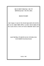
Figure 1: Organization of the mouse gastrointestinal tract Distinct regions of the gastrointestinal
tract of a mouse. The stomach is proximal to the small intestine which is separated into three
distinct regions; the duodenum, jejunum and ileum. The large intestine is also separated into three
distinct regions. The caecum is distal to the Ileum with the colon being proximal to the rectum
(Furness, 2012).
The GI tract is comprised of distinctly different cellular layers. The innermost layers (i.e., in close
proximity to the host organs) are the longitudinal muscle (LM), adjacent to the myenteric plexus
(MP), with the next layer being the circular muscle (CM) alongside the SMP muscularis mucosae (MM)
and the mucosa layer (Etherton et al.) (Figure 2). The intestinal mucosa is a single layer of epithelial
cells acting as the interface between the external gut environment and the internal environment of
the body. Epithelial cells absorb nutrients, water, and electrolytes, produce a variety of digestive
secretions and act as a physical barrier to potentially hazardous luminal substances. Enteroendocrine
cells (EECs) and other modified epithelial cell subtypes produce regulatory proteins that have local
paracrine or neurocrine effects. Paneth cells, for example, release antimicrobial mediators which
prevent invasions of potential pathogenic microbes into the epithelium (Allaire et al., 2018, Elphick
and Mahida, 2005). Similarly, mucus is secreted by goblet cells to lubricate the epithelial lining and
form a protective barrier against some microorganisms in the gut. The distribution of secretory cell
types varies by gut area and is intimately linked to the function of each gut region. Within the SMP is
a network of cells that coordinate the absorption, secretion, and immune activity of the mucosa. The
SMP consists of blood vessels, smooth muscle cells, neurons, and glial cells (Allaire et al., 2018).
6
The GI tract, in broad terms, protects the host from harmful substances that pass through the
intestinal epithelium. Neurons from the SMP mostly innervate the mucosa (Neunlist et al., 2013).
Immune cells and neurons work together to orchestrate inflammatory responses and evoke neuronal
signalling pathways that help the body eliminate infections. Neural activity regulates gut contractile
function as well as impacting permeability and secretion (e.g., of ions and water). Running the entire
length of the GI tract are three clearly defined smooth muscle layers, the CM, LM and the MM. As
their names suggests, the cells of the CM layer are oriented in a circular direction around the gut and
change gut diameter, while the cells in the outer LM layer are oriented along the gut's longitudinal
direction and change the gut length. Outside the lamina propria (Elphick and Mahida, 2005), the MM
is a thin layer of muscle that serves to separate it from the submucosa. Coordinated contractions and
relaxation of the CM and LM layers shape the complex motility contractions of the GI tract. These
patterns range from non-propulsive mixing movements to highly propulsive peristaltic contractions
(Costa et al., 2000, Furness, 2012, Furness et al., 2014). Embedded within the muscle layers of the GI
tract are two ganglionated plexus known as the SMP and the MP. Together these make up the ENS,
an intrinsic neural network that can regulate intestine activities independently of the CNS (Furness
et al., 2014). The ENS governs practically all gastrointestinal activities, including mucosal barrier
function, secretion, motility, and blood flow, for efficient food digestion and absorption. Although
the ENS can function independently of the CNS, it receives extrinsic innervation from the brain and
spinal cord to coordinate vital GI activities (Furness et al., 2014).
7
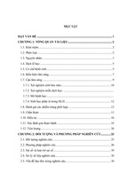
Figure 2: The organization of the enteric nervous system. In humans and large animals, the mucosa
is the intestine's exterior covering, which separates the lumen from the internal structures. The
submucosal and myenteric plexuses are the two ganglionic plexuses of the ENS. The sub-mucosal
plexus (SMP) runs between the mucosa layer and the circular muscle (CM) layer, and its ganglia are
divided into one or three layers. The SMP innervates the mucosa and regulates mucosal barrier
functions as secretion and permeability (Furness et al., 2014).
1.3 The enteric nervous system
The ENS is the largest division of the autonomic nervous system, with more than 100 million neurons
and 400 million neuron-supporting glial cells. This intricate neural system is crucial for maintaining
proper digestive function (Furness, 2012). Enteric nerve cells and glia are grouped together to form
ganglia in the ENS. A neural plexus is formed when ganglia are connected by nerve fiber bundles. The
SMP and the MP are two ganglionated plexuses in the ENS. The MP connects the LM and CM layers
of the external musculature, while the SMP connects the CM layer to the mucosa. The MP regulates
gut motility, while the SMP regulates mucosal barrier function, however, both plexuses work
together to ensure optimal GI functionality. Even though the ENS can perform functions independent
of the CNS, it is not autonomous. To govern local enteric reflexes, the integrated network of the ENS
and CNS mediates neuronal regulation of the GI system. Vagal nerve routes, the spinal thoracolumbar
8
spinal cord, and the pelvic pathway all play a role in neural communication between the CNS and the
ENS (Furness et al., 2013).
1.4 The submucosal plexus
The SMP is predominantly responsible for regulating water and electrolyte secretion along with
regulating localised blood flow (Furness, 2012). These processes are governed by a variety of enteric
neurons and elucidating the function of specific neuronal subtypes can offer insight into therapeutic
treatments for GI disorders. In the SMP of mice and guinea pigs, there are two pharmacologically and
neurochemically separate populations of neurons. Cholinergic neurons express choline
acetyltransferase (ChAT), the enzyme that synthesizes acetylcholine. Non-cholinergic neurons
possess vasoactive intestinal peptide (VIP) but lack the ability to express ChAT. Many VIP neurons in
humans and rats, on the other hand, express ChAT. SMP neurons express an array of neurochemicals
that provide them with a unique neurochemical code in addition to VIP and ChAT as two primary
neurotransmitters (Bornstein and Foong, 2018).
1.5 The myenteric plexus
The MP is located between the LM and CM layers and predominantly regulates GI motility including
peristaltic contractions of the smooth muscles to facilitate the transit of luminal contents (Furness,
2012). The MP comprises approximately 16 enteric neuronal subtypes which release a range of
excitatory (e.g. Acetylcholine and tachykinins) or inhibitory (i.e. nitric oxide, ATP (adenosine triphosphate)-like transmitters and VIP) neurotransmitters to contract or relax the intestinal smooth
muscle, respectively (Furness, 2012).
9
2.0 The gastrointestinal mucosal barrier
The intestinal epithelial wall of the human Gl tract covers an estimated 400 m2 of mucosal surface
area in an adult individual. Microorganisms, digestive enzymes and acids, digested food particles,
microbial by-products, and food-associated toxins all penetrate the mucus layer, which serves as
the GI tract's first line of defence. This mucus layer coats the surface of the GI tract, lubricates the
luminal contents, and acts as a physical barrier to bacteria and other antigenic chemicals present in
the lumen. The moist, nutrient-rich mucus layer next to the epithelial barrier of the GI tract is also
critical for intestinal homoeostasis because it contains a robust biofilm including both beneficial
and pathogenic bacteria species, reviewed by Herath and co-authors (Herath et al., 2020). The GI
tract is continually under attack by pathogens, medications, nutrients, and bacterial toxins. Not only
must the host distinguish between commensal bacteria and potential pathogens, but the host must
also prevent these species and secreted molecules from crossing the epithelial barrier while
allowing nutrients to be absorbed. Thus, the intestinal epithelium functions as a selective barrier to
luminal substances. This is accomplished in part by the innate epithelial defence system of the
mucosa, which operates via a responsive biological system composed of constitutive and inducible
mechanisms. Therefore, if the intestinal epithelial barrier's function is impaired, the host may be
more vulnerable to a variety of GI disorders. Consequently, this thesis will discuss how increased
paracellular permeability may be a possible explanation for why the NL3R451C mouse model exhibits
a dysfunctional epithelial barrier and how natural therapeutic treatments/agents such as, fasting
from solid foods, L-glutamine and caffeine supplementation may restore a compromised epithelial
barrier in (ASD).
10
- Xem thêm -