World J Microbiol Biotechnol (2012) 28:2029–2038
DOI 10.1007/s11274-012-1005-6
ORIGINAL PAPER
Comparison of the structural characterization and biological
activity of acidic polysaccharides from Cordyceps militaris
cultured with different media
Fengyao Wu • Hui Yan • Xiaoning Ma •
Junqiang Jia • Guozheng Zhang • Xijie Guo
Zhongzheng Gui
•
Received: 14 October 2011 / Accepted: 18 January 2012 / Published online: 18 March 2012
Ó Springer Science+Business Media B.V. 2012
Abstract Two acidic polysaccharide fractions, CM-jdCPS2 and CM-jd(Y)-CPS2, were isolated from the fruiting
bodies of cultured Cordyceps militaris grown on solid rice
medium and silkworm pupa, respectively, by hot-water
extraction, ethanol precipitation and fractionation using
ion-exchange column (DEAE-cellulose-52) and gel-filtration column (Sephadex G-100) chromatography. Their
structural characterizations were performed by gas chromatography and fourier-transform infrared spectroscopy.
Some differences existed between their structures, which
indicated that culture media could influence the structure of
polysaccharides of C. militaris. The antioxidant activities
of CM-jd-CPS2 and CM-jd(Y)-CPS2 were evaluated by
various methods in vitro. They had strong 2,2-diphenyl-1picrylhydrazyl radical-scavenging activity and ferrous ionchelating capacity, but moderate reducing power. The
antioxidant activities of CM-jd(Y)-CPS2 were slightly
higher than those of CM-jd-CPS2. These two acidic fractions were evaluated for proliferation of mouse splenocyte
activity in vitro. They both possessed does-dependent
mitogenic effects on mouse splenocytes, and could synergistically promote murine T- and B-lymphocytes induced
by Con A and LPS. CM-jd(Y)-CPS2 exhibited stronger
stimulatory activities upon immunomodulation than
CM-jd-CPS2. These results are beneficial for the
F. Wu Á H. Yan Á X. Ma Á J. Jia Á G. Zhang Á X. Guo Á
Z. Gui (&)
Jiangsu University of Science and Technology,
Zhenjiang 212018, China
e-mail:
[email protected]
J. Jia Á G. Zhang Á X. Guo Á Z. Gui
Sericultural Research Institute, Chinese Academy
of Agricultural Sciences, Zhenjiang 212018, China
interpretation of the connection between polysaccharide
structures and their biological activities.
Keywords Cordyceps militaris Á Media Á
Polysaccharide Á Structure Á Antioxidant Á Splenocyte
proliferation Á In vitro
Introduction
Cordyceps militaris is an entomogenous fungus that has
been used as a traditional Chinese medicine for centuries.
Studies have shown that it possesses similar constituents
and pharmacological efficacy to those of Cordyceps sinensis (Wei et al. 2004). These include immunomodulation
(Kuo et al. 2001; Kim et al. 2008), anti-tumor (Bok et al.
1999; Lin and Chiang 2008), and antioxidant effects
(Liu et al. 1997). In recent years, C. militaris has been
considered to be a suitable alternative to C. sinensis, which
is scarce due to problems with its artificial culture and
over-exploitation (Gui and Zhu 2008).
Polysaccharides from fungi used for medicinal purposes
have aroused wide interest because of their health benefits,
such as antioxidant capacity (Liu et al. 1997; Leung et al.
2009), and immunomodulatory activity (Wasser 2002).
Some mushroom polysaccharides such as lentinan and
schizophyllan have been applied or are in clinical trials as
immunomodifiers and adjuvant drugs for cancer therapies
(Zhang et al. 2007; Yang and Zhang 2009). It has been
reported that the bioactivities of polysaccharides are related
to their chemical composition, glycosidic linkages, conformation, molecular weight, and degree of branching
(Methacanon et al. 2005). As one of the major bioactive
constituents of C. militaris, several studies have been
123
2030
conducted to ascertain the structures and bioactivities of
different polysaccharides isolated from it.
In recent few years, investigators have explored different
types of media to artificially culture C. militaris. These
include silkworm pupa, solid rice medium, germinated
soybean medium, and soybean broth. Studies have shed light
on the structural characterizations, antioxidant activity and
immunomodulation of polysaccharides obtained from
fruiting bodies of cultured C. militaris grown on a particular
medium (Ohta et al. 2007; Leung et al. 2009). However, an
investigation comparing these properties between polysaccharides isolated from one strain of C. militaris grown on
different media has not been completed.
We investigated if there are differences with respect to
structural characterization, antioxidant activity and splenocyte proliferation between acidic polysaccharides obtained
from the fruiting bodies of cultured C. militaris grown on two
media. The results will be helpful for revealing connections
between the structure and function of polysaccharides.
World J Microbiol Biotechnol (2012) 28:2029–2038
collected as crude CM-jd-CPS and CM-jd(Y)-CPS (cultured on solid rice medium and silkworm pupa, respectively) and then lyophilized.
Isolation and purification of polysaccharides
Materials and methods
Each crude precipitate (100 mg), CM-jd-CPS and CMjd(Y)-CPS was dissolved in 10 mL distilled water, centrifuged at 8,000 rpm for 10 min at room temperature, and
loaded onto a DEAE-cellulose-52 column (2.6 9 30 cm).
After loading with sample, the column was eluted with
distilled water, and 0.1, 0.2 and 0.3 mol/L NaCl (each at
80 mL) at 1.0 mL/min. The eluate was collected at 4 min/
tube. This process was monitored by the phenol–sulfuric
acid method (Xu et al. 2005).
The resulting fraction (5 mg) was loaded onto a
Sephadex G-100 column (2.6 9 30 cm) for further purification, then eluted with distilled water. The flow rate was
0.5 mL/min. Consequently, the homogeneous fractions,
CM-jd-CPS2 and CM-jd(Y)-CPS2, were obtained and
lyophilized. They were used in the subsequent structural
and bioactive studies.
Fungal strains and materials
Ultraviolet spectroscopy of polysaccharides
The C. militaris strain used was CM-jd, which was conserved by our research team. Fresh fruiting bodies grown
on a solid rice medium and silkworm pupa were obtained.
2,2-Diphenyl-1-picrylhydrazyl (DPPH), ascorbic acid
(vitamin C), ferrozine, potassium ferricyanide, ferrous chloride
and ferric chloride were purchased from Bio Basic Inc (Toronto, Canada). RPMI-1640 medium, fetal calf serum (FCS)
and dimethylsulfoxide (DMSO) were from Gibco Laboratories
(Gaithersburg, MD, USA). Concanavalin A (Con A), lipopolysaccharide (LPS) and 3-(4,5-dimethylthiazol-2-yl)-2,5diphenyltetrazolium bromide (MTT) were purchased from
Sigma-Aldrich (St Louis, MO, USA). All other reagents were
obtained from Sinopharm Chemical Reagent Co. Ltd. (Beijing, China). All reagents were of analytical grade.
CM-jd-CPS2 and CM-jd(Y)-CPS2 were dissolved in distilled water to 0.1 mg/mL. They were scanned with a UV
spectrophotometer (UV-2450, Shimadzu, Beijing, China)
at wavelengths from 800 to 200 nm.
Extraction of polysaccharides
The dried powder of C. militaris fruiting bodies (100 g)
cultured on solid rice medium and silkworm pupa were
defatted with ethanol for 10 h twice at 70°C, and exhaustively extracted twice with 20 volumes of water at 80°C,
each time for 10 h. Extracts were concentrated under
reduced pressure to 100 mL, deproteinated by the Sevag
method (Wu et al. 2011) and dialyzed against distilled
water for 4 days to remove low-molecular-weight compounds. Crude polysaccharides were obtained through
precipitation with ethanol to a final concentration of ethanol of 90%, and left overnight at 4°C. Precipitates were
123
Gas chromatography (GC) analyses
Derivatives of acid hydrolytic products from CM-jd-CPS2
and CM-jd(Y)-CPS2 were analyzed by GC to identify their
monosaccharide components.
Each fraction (10 mg) was placed in an ampoule. It was
hydrolyzed with 3 mL of 4 mol/L trifluoroacetic acid
(TFA) at 115°C for 12 h. The ampoule was sealed under a
nitrogen atmosphere. The acidolysis solution was dried
with a stream of N2 at 65°C in a water bath. The solid
residual was re-dissolved in methanol (1 mL) and then
distilled at 65°C with a stream of N2. This process was
repeated thrice to remove the acid.
The solid hydrolysate was mixed with hydroxylamine
hydrochloride (8 mg) and inositol (10 mg) as internal standard. They were then re-dissolved in pyridine (0.5 mL) and
allowed to react with shaking at 95°C for 30 min. The sample
was then cooled to room temperature. Acetic anhydride
(0.5 mL) was added to continue the acetylation reaction at
95°C for 30 min. Upon reaction completion, the solution was
mixed with methanol (2 mL), and dried with a stream of N2.
The derivatives were dissolved in chloroform (1 mL) and
analyzed by GC. The derivation of mixed standard
World J Microbiol Biotechnol (2012) 28:2029–2038
monosaccharides (rhamnose, arabinose, xylose, mannose,
glucose, galactose) was operated using the same method as
described above.
GC was undertaken on an Agilent 6820 Gas Chromatography system (Agilent Technologies, Santa Clara, CA,
USA) equipped with a flame ionization detector (FID),
through a fused-silica capillary column (0.23 mm 9 30 m).
N2 was used as the carrier gas, and H2 as the burning gas.
The sample injection volume was 1 lL at a N2 flow of
50 mL/min at a split ratio of 50:1. The injection temperature
and FID detector were controlled at 230°C. The column
temperature was first fixed at 130°C for 20 min, increased to
190°C at 5°C/min, and maintained for 20 min, then
increased to 230°C at 5°C/min, and fixed for 10 min.
Infrared (IR) analyses
IR spectrometry of CM-jd-CPS2 and CM-jd(Y)-CPS2 was
done in the 4,000–400 cm-1 wavenumber range. The dried
sample (1–2 mg) was pressed into KBr (100–200 mg) disks,
and then scanned with a fourier-transform infrared (FTIR)
spectrometer (Tensor 27; Bruker, Billerica, MA, USA).
2031
incubated at 50°C for 20 min. Then, 2 mL of trichloroacetic
acid (10%, w/v) was added to the mixture to terminate the
reaction, and centrifuged at 5,000 rpm for 10 min at room
temperature. The supernatant (2.5 mL) was mixed with distilled water (2.5 mL) and 0.5 mL ferric chloride (0.1%, w/v)
and allowed to stand for 10 min. The absorbance was measured
at 700 nm. Vc was used as the positive control. Increased
absorbance of the reaction mixture indicated increased reducing power.
Ferrous ion-chelating capacity assay
Ferrous ion-chelating capacity was determined according to
the method of Decker and Welch (1990) with some modification. The reaction mixture, containing 3 mL of each sample with different concentrations (2, 4, 6 and 8 mg/mL),
0.05 mL ferrous chloride solution (2 mmol/L), and 0.2 mL
of ferrozine solution (5 mmol/L), was shaken vigorously and
incubated at room temperature for 10 min. The absorbance of
the mixture was measured at 562 nm. Ethylenediamine tetraacetic acid disodium salt (EDTA-2Na) was used as the
positive control. The ferrous ion-chelating capacity of the
sample was calculated using the following formula:
In vitro antioxidant assay
Chelating capacity ð%Þ ¼
2,2-diphenyl-1-picrylhydrazyl (DPPH) radical-scavenging
assay
DPPH radical-scavenging capacity was assayed according
to the method of Luo et al. (2009). with some modifications. Each sample (2 mL) at different concentrations (2, 4,
6 and 8 mg/mL) was mixed with a solution of 0.04 mg/mL
DPPHÁ (2 mL) in ethanol. The mixture was shaken vigorously and allowed to stand at room temperature for 30 min.
Then, it was centrifuged at 5,000 rpm for 10 min at room
temperature. The absorbance of the supernatant was measured at 517 nm. Vitamin C (Vc) was used as the positive
control. DPPH radical-scavenging capacity was calculated
using the following formula:
Scavenging capacity ð%Þ ¼
1 À ðA1 À A2 Þ
 100
A0
where A0 is the absorbance of the control (ethanol instead
of sample), A1 is the absorbance of the sample, and A2 is
the absorbance of the blank (which was obtained by
replacing the DPPHÁ ethanol solution with ethanol).
A0 À ðA1 À A2 Þ
 100
A0
where A0 is the absorbance of the control (water instead of
sample), A1 is the absorbance of the sample, and A2 is the
absorbance of the blank (which was obtained by replacing
the FeCl2 solution with distilled water).
Measurement of immunomodulatory activity
Preparation of mouse spleen cells
Male Kunming mice were killed by cervical dislocation. The
spleens were removed, minced and washed through a sterilized copper mesh (200 mesh) by RPMI-1640 (5 mL) to obtain
a suspension of single spleen cells. The suspension was centrifuged at 1,500 rpm for 5 min at 4°C to obtain precipitated
cells. Erythrocytes in precipitated cells were lysed with TrisNH4Cl solution (0.14 mol/L NH4Cl and 20 mmol/L Tris) for
2–3 min. The lysed solution was centrifuged at above condition, washed twice with RPMI-1640 medium, and adjusted
to 5 9 106 cells/mL in the RPMI-1640 medium supplemented with 10% of FBS (Fetal bovine serum), penicillin
(100 U/mL) and streptomycin (100 lg/mL).
Reducing power assay
Assay of splenocyte proliferation
Reducing power was determined by the method of Tsais et al.
(2006). Briefly, 1 mL of different concentrations of samples
(2, 4, 6 and 8 mg/mL) in phosphate buffer (0.2 mol/L, pH 6.6)
was mixed with 2 mL potassium ferricyanide (1%, w/v), and
Isolated splenocytes (100 lL/well) were seeded onto a
96-well plate in the presence or absence of three concentrations (50, 100 and 200 lg/mL) of each sample (100 lL).
123
2032
World J Microbiol Biotechnol (2012) 28:2029–2038
Con A (5 lg/mL) was used as the positive control. After
incubation for 48 h at 37°C in a humidified incubator
containing 5% CO2, MTT solution (5 mg/mL; 20 lL/well)
was added and the plate incubated for a further 4 h. After
removing MTT by centrifugation at 1,000 rpm for 5 min at
4°C, the formazan precipitate was solubilized in DMSO
(100 lL/well). The absorption of each well was measured
using an enzyme-linked immunosorbent assay (ELISA)
reader (EL310, Bio-TKE Instruments, Winooski, VT,
USA) at 570 nm.
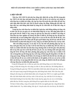
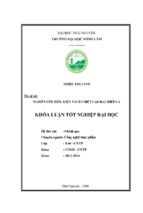
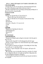
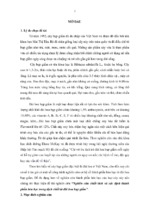
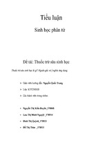
CPS2. These acidic fractions were collected and applied to
a Sephadex G-100 gel filtration column, each giving a
single elution peak in Fig. 1c, d, which elucidated the
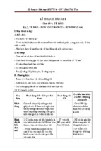
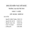
homogeneity of the fraction. The UV absorption spectra
(Fig. 2) of CM-jd-CPS2 and CM-jd(Y)-CPS2 showed no
absorption at 260 and 280 nm, indicating that nucleic acids
and proteins were absent in these polysaccharides.
Assay of Con A- or LPS-induced splenocyte proliferation
GC analyses of CM-jd-CPS2 and CM-jd(Y)-CPS2
Splenocytes (prepared by following the procedure described above) were seeded onto a 96-well plate (100 lL/
well), and mixed with serial dilutions of each sample (50,
100 and 200 lg/mL). Cells were incubated in the presence
of Con A (5 lg/mL) or LPS (10 lg/mL) for 48 h in a
humidified incubator containing 5% CO2 at 37°C. MTT
solution (5 mg/mL) was added to each well (20 lL/well),
and the plate incubated at 37°C for a further 4 h. The
medium was removed and the formazan crystals formed
dissolved by adding DMSO (100 lL/well). The absorption
of each well was measured using an ELISA reader at
570 nm. Control experiments were undertaken without
polysaccharide samples.
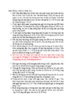
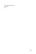
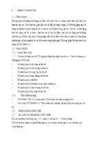
The alditol acetates derived from the acetylation of CM-jdCPS2, CM-jd(Y)-CPS2 hydrolysate and standard monosaccharide were measured by GC (Fig. 3). Mixtures of
monosaccharide and inositol were completely separated
(Fig. 3a). The peaks emerged in the order: rhamnose,
arabinose, xylose, mannose, glucose, galactose and inositol. Figure 3b, c show that three types of monosaccharide
(mannose, glucose and galactose) were identified in the
hydrolysate of CM-jd-CPS2 and CM-jd(Y)-CPS2 on the
basis of the retention time and correction factor of standard
monosaccharides. According to the peak area, the mole
ratio of mannose:glucose:galactose in CM-jd-CPS2 was
1.52:8.53:1.00, and in CM-jd(Y)-CPS was 3.11:1.00:2.12.
These two fractions comprised the same kinds of
monosaccharide, but there was a large difference in the
mole ratio of each type of monosaccharide in their
polysaccharides. The content of glucose was highest in
CM-jd-CPS, but that of mannose and galactose much
lower. Conversely, mannose comprised the highest content in CM-jd(Y)-CPS, but glucose content was the
lowest.
Statistical analyses
All treatments and assays were carried out in triplicate for
three separate experiments. Values are mean ± SD. The
statistical significance was analyzed by Student’s t test and
regression analysis and the data were fitted by using the
Expert Design 7.1.3 for Windows software (SPSS Inc.,
USA).
Comparison of the structural characterizations
of CM-jd-CPS2 and CM-jd(Y)-CPS2
IR analyses of CM-jd-CPS2 and CM-jd(Y)-CPS2
Results
Isolation and purification of CM-jd-CPS and CM-jd(Y)CPS
Through hot-water extraction and ethanol precipitation, the
crude polysaccharides CM-jd-CPS and CM-jd(Y)-CPS
were isolated from the fruiting bodies of C. militaris cultured with solid rice medium and silkworm pupa, respectively. Figure 1a, b show the elution profiles of
deproteinized CM-jd-CPS and CM-jd(Y)-CPS on a DEAEcellulose-52 ion-exchange column. In each profile, the first
peak (which was eluted with distilled water) was ascribed
to a neutral polysaccharide fraction. The main peak subsequently eluted with NaCl solution was an acidic polysaccharide fraction termed CM-jd-CPS2 and CM-jd(Y)-
123
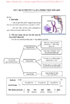
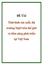
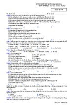
In the IR spectrum of CM-jd-CPS2 (Fig. 4a), the largest
absorption band (at 3,396 cm-1), was ascribed to the
stretching of the hydroxyl group and the hydrogen bond
within or between the molecules. The peak at 2,927 cm-1
was attributed to the C–H stretching band of the saccharide, and the weak peaks between 1,400 and 1,200 cm-1
ascribed to the C–H bending vibration. The bands at 1,651
and 1,541 cm-1 were assigned to the stretching vibration
of the C=O bond and the bending vibration of the N–H
bond, which suggested the presence of an acetamido group.
The two absorption bands near 1,240 and 850 cm-1
resembled the stretching band of S=O and C–O–S,
respectively, which indicated the existence of –O–SO3.
There was a group of strong absorption peaks from 1,200 to
950 cm-1 which could be attributed to the ether linkage
(C–O–C) and the hydroxyl present in the pyranose ring.
World J Microbiol Biotechnol (2012) 28:2029–2038
2033
300
ntration of polysaccharide (m
mg/ml)
Concen
Conce
entration of polysaccharide (
(mg/ml)
350
A
300
250
200
150
100
50
B
250
200
150
100
50
0
0
0
10
20
30
40
50
60
70
80
0
90
10
C
Concentration of polysaccharide (mg/ml)
f
Concentration of polysaccharide (mg/ml)
30
40
50
60
70
80
90
18
40
35
30
25
20
15
10
5
0
20
No. tube
No. tube
0 2 4 6 8 10 12 14 16 18 20 22 24 26 28 30 32 34 36 38 40 42 44 46 48 50
No. tube
16
D
14
12
10
8
6
4
2
0
0 2 4 6 8 10 12 14 16 18 20 22 24 26 28 30 32 34 36 38 40 42 44 46 48 50
No. tube
Fig. 1 Elution profiles of polysaccharides extracted from cultured
C. militaris by column chromatography. a and b Ion exchange
chromatogram of crude polysaccharides, CM-jd-CPS and CM-jd(Y)-
CPS, on a DEAE-cellulose-52 column. c and d Gel filtration
chromatogram of acidic polysaccharide fractions, CM-jd-CPS2 and
CM-jd(Y)-CPS2, on a Sephadex G-100 column
Fig. 2 Ultraviolet absorption curve of CM-jd-CPS2 and CM-jd(Y)CPS2
The peak at 761 cm-1 was the symmetric ring stretching
of pyranose, which also implied that the monosaccharide
in CM-jd-CPS2 was a pyranose. The presence of the
a-glycosidic linkage was proven by the C–H bending
vibration at 850 cm-1. The absorption peaks at 931 and
761 cm-1 suggested the existence of a-D-glucopyranose
(a-D-Glcp).
CM-jd(Y)-CPS2 also possessed the characteristic
absorption peaks of a saccharide at 3,600–3,200,
3,000–2,800 and 1,400–1,200 cm-1 (Fig. 4b). The bands at
1,652 and 1,541 cm-1 indicated an acetamido group. The
absorption band at 1,220 cm-1 was ascribed to the bending
vibration of O–H in a carboxy group (–COOH). The bands
of a C–O stretching vibration in the carboxy group at
1,440–1,395 cm-1 further proved the presence of a carboxy group in CM-d(Y)-CPS2. The group of strong
absorption peaks from 1,200 to 950 cm-1 suggested that
the monosaccharide in CM-jd(Y)-CPS2 was a pyranose.
123
2034
Fig. 3 GC profiles of standard monosaccharide (a), CM-jd-CPS2
(b) and CM-jd(Y)-CPS2 (c). a Peak identity: 1 Rhamnose (rt: 30.897);
2 Arabinose (rt: 31.336); 3 Xylose (rt: 31.803); 4 Mannose (rt: 40.963);
5 Glucose (rt: 41.530); 6 Galactose (rt: 42.978); 7 Inositol as an internal
World J Microbiol Biotechnol (2012) 28:2029–2038
standard. b Peak identity: 1 Mannose (rt: 41.195); 2 Glucose (rt:
41.816); 3 Galactose (rt: 43.213); 4 Inositol as an internal standard.
c Peak identity: 1 Mannose (rt: 40.866); 2 Glucose (rt: 41.376); 3
Galactose (rt: 42.830); 4 Inositol as an internal standard
Fig. 4 IR spectrum of CM-jd-CPS2 (a) and CM-jd(Y)-CPS2 (b)
Unlike CM-jd-CPS2, there was b-glycosidic linkage in
CM-jd(Y)-CPS2 which was ensured by a C–H bending
vibration at 893 cm-1(Wang et al. 2011).
123
From the results described above, it could be concluded that
CM-jd-CPS2 was a type of sulfated polysaccharide containing
an acetamido group. The monosaccharide in CM-jd-CPS2 was
World J Microbiol Biotechnol (2012) 28:2029–2038
2035
a pyranose, which was connected by an a-glycosidic linkage.
CM-jd(Y)-CPS2 was a type of carboxylated polysaccharide
containing an acetamido group. The monosaccharide was a
pyranose, which was linked by a b-glycosidic linkage.
for both of them was much lower than that of Vc. This
finding suggested that CM-jd-CPS2 and CM-jd(Y)-CPS2
had moderate reducing power.
Ferrous ion-chelating activity of CM-jd-CPS2
and CM-jd(Y)-CPS
In vitro antioxidant activities of CM-jd-CPS2
and CM-jd(Y)-CPS2
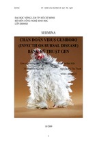
In the present study, the ferrous ion-chelating capacity of
antioxidants was detected by inhibiting the formation of
red-colored ferrozine–Fe2? complexes. At 8 mg/mL, the
ferrous ion-chelating activities of CM-jd-CPS2 and CMjd(Y)-CPS2 were 72 and 89%, respectively, and both
activities were concentration-related (Fig. 5c). However,
overall, CM-jd(Y)-CPS2 possessed higher ferrous ion-chelating activity than CM-jd-CPS2 with, on average, &20%
higher activity at each concentration. The positive control
EDTA-2Na had excellent ferrous ion-chelating activities
(100% at each concentration tested). CM-jd(Y)-CPS2
exhibited strong Fe2?-chelating activities at 4, 6 and 8 mg/
mL, but that of CM-jd-CPS2 was comparatively weak.
DPPH radical-scavenging activity of CM-jd-CPS2
and CM-jd(Y)-CPS2
CM-jd-CPS2 and CM-jd(Y)-CPS2 exerted concentrationdependent DPPHÁ-scavenging activity (Fig. 5a). At 8 mg/
mL, DPPHÁ-scavenging activity for CM-jd-CPS2 and CMjd(Y)-CPS2 reached &94%. However, at a low concentration (2 mg/mL), the scavenging activity of CM-jd-CPS2
was higher than that of CM-jd(Y)-CPS2. Compared with
the positive control (Vc), the scavenging activity of these
two fractions were slightly lower than Vc at each concentration. These results indicated that CM-jd-CPS2 and
CM-jd(Y)-CPS2 had strong DPPHÁ-scavenging activities.
In vitro immunomodulatory activity of CM-jd-CPS2
and CM-jd(Y)-CPS2
Reducing power of CM-jd-CPS2 and CM-jd(Y)-CPS2
Effect of CM-jd-CPS2 and CM-jd(Y)-CPS2 on splenocyte
proliferation
Samples showed a dose-dependent reducing capacity
(Fig. 5b). At 8 mg/mL, the reducing power of CM-jd-CPS2
was 0.985, for CM-jd(Y)-CPS it was 1.214, and for Vc it
was 2.108. The reducing power of CM-jd(Y)-CPS2 was
higher than that for CM-jd-CPS2, but the reducing power
B
100
95
Absorbency
Scavenging capacity (%)
A
90
85
CM-jd-CPS2
CM-jd(Y)-CPS2
j ( )
Vc
80
75
70
2
4
6
8
Concentration (mg/ml)
2.2
2
1.8
1.6
1.4
1.2
1
0.8
0.6
0.4
0.2
0
CM-jd-CPS2
CM jd CPS2
CM-jd(Y)-CPS2
Vc
2
4
6
8
Concentration (mg/ml)
C 120
Chelating capacity (%)
Fig. 5 The antioxidant
activities of CM-jd-CPS2 and
CM-jd(Y)-CPS2. a DPPH
radical scavenging activities
with Vc as the positive control.
b The reducing power assay
with Vc as the positive control.
c The ferrous ion chelating
capacity with EDTA-2Na as the
positive control. Values are
represented as mean ± SD
(n = 3)
A colorimetric assay using MTT for cell proliferation was
carried out to evaluate the effect of CM-jd-CPS2 and
100
80
60
CM-jd-CPS2
CM-jd(Y)-CPS2
40
EDTA
20
0 2
4
6
8
Concentration (mg/ml)
123
2036
World J Microbiol Biotechnol (2012) 28:2029–2038
Table 1 Effect of CM-jd-CPS2 and CM-jd(Y)-CPS2 on mouse
splenocyte proliferation
Preparation
Dose (lg/mL)
Control
A570
–
5
Positive control (Con A)
CM-jd-CPS
0.256 ± 0.001
0.376 ± 0.007
50
0.265 ± 0.003
100
0.279 ± 0.006a
200
0.293 ± 0.006a
50
0.283 ± 0.008a
100
200
0.306 ± 0.003b
0.362 ± 0.002b
CM-jd(Y)-CPS
a
P \ 0.01 when compared with the control group
Discussion
P \ 0.05 when compared with the control group
b
compared with the Con A control group (P \ 0.01). The
stimulatory effects of CM-jd-CPS2 and CM-jd(Y)-CPS2
upon lymphocyte proliferation induced by LPS were nearly
identical, and were both significantly increased (P \ 0.01)
at 200 lg/mL. These results suggested that CM-jd-CPS2
and CM-jd(Y)-CPS2 could dose-dependently promote
murine T- and B-cell proliferation induced by specific
mitogens, whereas CM-jd(Y)-CPS2 possessed a stronger
stimulatory activity to T cells than CM-jd-CPS2.
Table 2 Effects of CM-jd-CPS2 and CM-jd(Y)-CPS2 on Con A or
LPS induced mouse splenocyte proliferation
Preparation
Control (Con A)
Dose (lg/mL)
5
Control (LPS)
10
CM-jd-CPS
50
Con A (A570)
LPS(A570)
0.376 ± 0.007
–
–
0.390 ± 0.007
0.380 ± 0.005
0.398 ± 0.009c
a
0.415 ± 0.004c
200
a
0.407 ± 0.004
0.425 ± 0.003d
50
0.403 ± 0.006a
0.403 ± 0.006c
100
200
b
0.415 ± 0.007c
0.430 ± 0.001d
100
CM-jd(Y)-CPS
a
0.396 ± 0.002
0.430 ± 0.005
0.440 ± 0.003b
P \ 0.05 when compared with the Con A control group
b
P \ 0.01 when compared with the Con A control group
c
P \ 0.05 when compared with the LPS control group
d
P \ 0.01 when compared with the LPS control group
CM-jd(Y)-CPS2 upon splenocyte proliferation (Table 1). In
comparison with the control group (without sample treatment), promoting effects increased with increasing sample
concentration (50–200 lg/mL). CM-jd(Y)-CPS2 produced
statistically significant promotion (P \ 0.01) of proliferation
of mouse splenocytes at 200 lg/mL, which was close to that
of the positive control group (Con A). CM-jd-CPS2 showed
weaker stimulating activity than that of CM-jd(Y)-CPS2.
Effect of CM-jd-CPS2 and CM-jd(Y)-CPS2 on
Con A- or LPS-induced splenocyte proliferation
The effects of CM-jd-CPS2 and CM-jd(Y)-CPS2 on lymphocyte proliferation induced by Con A or LPS were
investigated. The stimulatory effects of the two samples
upon proliferation were higher with increasing doses
(Table 2). The proliferation of lymphocytes induced by
Con A was promoted by &17% by CM-jd(Y)-CPS2 at
200 lg/mL, which was a statistically significant promotion
123
Cultivated fruiting bodies of C. militaris have been sold as
drug materials and healthfood products in China and South
East Asia in recent years. With the growing demand of its
fruiting bodies, culturing it with silkworm pupa was not
sufficient to match market requirements. Hence, solid rice
medium (which is relatively plentiful and not affected by
seasonal changes) was used to cultivate the fruiting bodies
of C. militaris on a large scale. However, there is no report
comparing the bioactive constituents contained in fruiting
bodies cultured by these two media. As one of its main
components, polysaccharides have attracted wide attention.
Polysaccharides have the greatest potential for structural
variability to carry biological information, so it is important to determine their structure and bioactivity.
In the last few years, structural characterizations of
several polysaccarides obtained from cultured C. militaris
have been reported. An acidic polysaccharide isolated from
C. militaris grown on germinated soybeans was found to be
composed of galactose, arabinose, xylose, rhamnose and
galacturonic acid (Ohta et al. 2007). CPS-2 was isolated
from cultured C. militaris, and primarily comprised
rhamnose, glucose and galactose in a molar ratio of
1:4.46:2.43 (Yu et al. 2004). Lee et al. (2010) reported the
structural properties of CPSN Fr II, which was obtained
from the cultured mycelia of C. militaris. CPSN Fr II was a
1,6-branched-glucogalactomannan with a b-linkage and
random coil conformation. The interpretation of structural
differences between the results described above may be
because they were different strains of C. militaris. Otherwise, different culture media (silkworm pupa, solid rice
medium, and broth) may contribute to the differences
between the structures of the polysaccharide. In the present
study, two acidic polysaccharides, CM-jd-CPS2 and CMjd(Y)-CPS2, were extracted from the fruiting bodies of the
same strain of C. militaris cultivated on solid rice medium
and silkworm pupa, respectively. Structure elucidation
showed that CM-jd-CPS2 and CM-jd(Y)-CPS2 comprised
mannose, glucose and galactose, but with different proportions. CM-jd-CPS2 had a large proportion of glucose,
whereas CM-jd(Y)-CPS2 had a large proportion of
World J Microbiol Biotechnol (2012) 28:2029–2038
mannose. The IR spectrum revealed that the monosaccharide within them was a pyranose, that CM-jd-CPS2 was
connected by an a-glycosidic linkage, and that CM-jd(Y)CPS2 was connected by a b-glycosidic linkage. In addition,
CM-jd-CPS2 was a type of sulfated acidic polysaccharide
containing an acetamido group, whereas CM-jd(Y)-CPS2
was a kind of carboxylated polysaccharide. These results
indicated that the differences between their structures were
due to different culture media. Hence, we showed that
culture media could influence the structure of polysaccharides of C. militaris.
Polysaccharides and their derivatives are emerging as
new options for combating oxidative stress-mediated disorders (Huang et al. 2009). The antioxidant properties of
polysaccharides are very relevant to their health-protecting
and anti-cancer functions. These two acidic fractions could
efficiently scavenge the stable free radical DPPHÁ. This was
attributed to their electron-transfer or hydrogen-donating
ability. It has been suggested that the hydroxyl (–OH)
group in polysaccharides can donate electrons to reduce the
radicals to a more stable form or react with the free radicals
to terminate the radical chain reaction (Leung et al. 2009).
CM-jd-CPS2 and CM-jd(Y)-CPS2 showed moderate
reducing power. This may have been due to the –OH group
and certain reducing groups in their structures. The presence of reductants is associated with reducing power.
Reductants have been shown to exert antioxidant actions
by breaking the free-radical chain by donating a hydrogen
atom (Zhang et al. 2010a, b). The results of the ferrous ionchelating capacity assay showed that CM-jd-CPS2 and
CM-jd(Y)-CPS2 exhibited strong Fe2?-chelating activities
at high concentrations. It has been demonstrated that
compounds with metal-chelating activities usually contain
two or more of the following functional groups: –OH, –SH,
–COOH, –PO3H2, –C=O, –NR2, –S– and –O– (Yuan et al.
2005). Accordingly, the ferrous ion-chelating capacities of
the two acidic polysaccharides were partially accounted for
by the presence of –OH, C=O, –S– and –O– groups in their
structure. Moreover, it has been reported that the antioxidant capacity of polysaccharides is also strongly dependent
upon the type and organization of the monosaccharide, the
linkage pattern of the main chain (a or b) and the branching
configuration (Liu et al. 2007). This could explain the
differences of reducing power and ferrous ion-chelating
capacity between CM-jd-CPS2 and CM-jd(Y)-CPS2.
The therapeutic effects of C. militaris have been shown
to be mediated through reinforcement of the immune system (Won and Park 2005). The spleen is one of the major
immune organs, and contains T-and B-lymphocytes.
Splenocyte proliferation is related to the improvement of
immunity of T- and B-lymphocytes (Qiao et al. 2010). In
the present study, CM-jd-CPS2 and CM-jd(Y)-CPS2 had
direct mitogenic effects on mouse splenocytes, which could
2037
strengthen the immunological response. CM-jd(Y)-CPS2
showed mitogenic effect, but it was not comparable than
CM-jd-CPS2. These acidic polysaccharides affected the
Con A- and LPS-induced splenocyte proliferation.
Administration of CM-jd(Y)-CPS2 at high concentrations
was found to significantly increase the proliferation of
splenocytes. CM-jd-CPS2 was the one to have mitogenic
effect but this effect was not comparable with CM-jd(Y)CPS2. The two acidic fractions strongly increased proliferation of splenocytes with LPS at 200 lg/mL. It is known
that the combination of polysaccharides and Con A can
significantly promote the proliferation of T-lymphocytes
(Yuan et al. 2005; Zhao et al. 2006; Zhang et al. 2010a, b).
These results demonstrated that CM-jd-CPS2 and
CM-jd(Y)-CPS2 could synergistically promote the induction of murine T- and B-lymphocytes by specific mitogens.
CM-jd(Y)-CPS2, which was obtained from silkworm pupacultivated C. militaris, possessed stronger stimulatory
activity on immunomodulation than CM-jd-CPS2. It has
been reported that many factors influence the activities of
polysaccharides, including monosaccharide composition,
glycosyl residues, chain conformation and molecular mass
(Bao et al. 2001; Huang et al. 2007). Consequently, the
different immunomodulatory activities of the two acidic
polysaccharides may have been due to differences in their
structures. Also, some researches suggested that acidic
polysaccharides are likely to be more bioactive than neutral
polysaccharides because the acidic groups form associations with target biomolecules through electronic interactions (Tadera et al. 2003). It was found that the acidic
polysaccharide from Tanacetum vulgare L. showed higher
immuno-activities than the neutral polysaccharide fraction
(Won and Park 2005; Xie et al. 2007).
The fundamental findings in the present study are beneficial for interpretation of the connection between polysaccharide structures and their biological activities.
However, because of the complex structures of bioactive
polysaccharides, it is difficult to elucidate their chemical
properties and relationships between their structure and
activity. Therefore, great efforts should be devoted to
reveal the structure–activity relationship of polysaccharides isolated from C. militaris in further studies.
Acknowledgments This work was supported by the Key Technologies R&D Program of China grant No. 2011BAD33B04, and the
Key Technologies R&D Program of Jiangsu province grant No.
BE2011389.
References
Bao XF, Liu CP, Fang JN, Li XY (2001) Structural and immunological studies of a major polysaccharide from spores of
Ganoderma lucidum (Fr.) Karst. Carbohydr Res 332:67–74
123
2038
Bok JW, Lermer L, Chilton J, Klingeman HG (1999) Antitumor
sterols from the mycelia of Cordyceps sinensis. Phytochemistry
51:891–898
Decker EA, Welch B (1990) Role of ferritin as a lipid oxidation
catalyst in muscle food. J Agric Food Chem 38:674–677
Gui ZZ, Zhu YH (2008) Advance on cultivation, bioactive compound
and pharmacological mechanism of Cordyceps militaris. Sci Seri
34:178–184
Huang QL, Jin Y, Zhang LN, Cheung PCK, Kennedy JF (2007)
Structure, molecular size and antitumor activities of polysaccharides from Poria cocos mycelia produced in fermenter.
Carbohydr polym 70:324–333
Huang XJ, Li Q, Li HG, Guo LJ (2009) Neuroprotective and
antioxidative effect of cactus polysaccharides in vivo and in
vitro. Cell Mol Neurobiol. doi:10.1007/s10571-009-9417-z
Kim CS, Lee SY, Cho SH, Ko YM, Kim BH, Kim HJ, Park JC, Kim
DK, Ahn H, Kim BO, Lim SH, Chun HS, Kim DK (2008)
Cordyceps militaris induces the IL-18 expression via its
promoter activation for IFN-c production. J Ethnopharmacol
120:366–371
Kuo YC, Tsai WJ, Wang JY, Chang SC, Lim CY (2001) Regulation
of bronchoalveolar lavage fluids cell function by the immunomodulatory agents from Cordyceps sinensis. Life Sci 68:67–82
Lee JS, Kwon JS, Won DP, Lee KE, Shin WC, Hong EK (2010)
Study on macrophage activation and structural characteristics of
purified polysaccharide from the liquid culture broth of Cordyceps militaris. Carbohydr Polym 82:982–988
Leung PH, Zhao S, Ho KP, Wu JY (2009) Chemical properties and
antioxidant activity of exopolysaccharides from mycelial culture
of Cordyceps sinensis fungus Cs-HK1. Food Chem 114:
1251–1256
Lin YW, Chiang BH (2008) Anti-tumor activity of the fermentation
broth of Cordyceps militaris cultured in the edium of Radix
astragali. Process Biochem 43:244–250
Liu F, Ooi VEC, Chang ST (1997) Free radical scavenging activities
of mushroom polysaccharide extracts. Life Sci 60:763–771
Liu CH, Wang CH, Xu ZL, Wang Y (2007) Isolation, chemical
characterization and antioxidant activities of two polysaccharides from the gel and the skin of Aloe barbadensis Miller
irrigated with sea water. Process Biochem 42:961–970
Luo W, Zhao MM, Yang B, Shen GL, Rao GH (2009) Identification
of bioactive compounds in Phyllenthus emblica L. fruit and their
free radical scavenging activities. Food Chem 114:499–504
Methacanon P, Madla S, Kirtikara K, Prasitsil M (2005) Structural
elucidation of bioactive fungi-derived polymers. Carbohydr
Polym 60:199–203
Ohta Y, Lee JB, Hayashi K, Fujita A, Park DK, Hayashi T (2007)
In vivo anti-influenza virus activity of an immunomodulatory
acidic polysaccharide isolated from Cordyceps militaris grown
on germinated soybeans. J Agric Food Chem 55:10194–10199
Qiao DL, Luo JG, Ke CL, Sun Y, Ye H, Zeng XX (2010)
Immunostimulatory activity of the polysaccharides from Hyriopsis Cumingii. Int J Biol Macromol 47:676–680
123
World J Microbiol Biotechnol (2012) 28:2029–2038
Tadera K, Minamiy Y, Chohchi M (2003) Interaction between acidic
polysaccharides and proteins. Biosci Biotech Biochem 67:
1840–1843
Tsais Y, Huang SJ, Mau JL (2006) Antioxidant properties of hot
water extracts from Agrocybe cylindracea. Food Chem
98:670–677
Wang ZM, Peng X, Lee KD, Tang JC, Cheung PCK, Wu JY (2011)
Structural characterization and immunomodulatory property of
an acidic polysaccharide from mycelial culture of Cordyceps
sinensis fungus Cs-HK1. Food Chem 125:637–643
Wasser SP (2002) Medicinal mushrooms as a source of antitumor and
immunomodulating polysaccharides. Appl Microbiol Biotechnol
60:258–274
Wei HP, Xiao B, Hu KZ (2004) Pharmaceutical values of Cordyceps
militaris. J Chin Med Mater 27:215–217
Won SY, Park EH (2005) Anti-inflammatory and related pharmacological activities of cultured mycelia and fruiting bodies of
Cordyceps militaris. J Ethnopharmacol 96:555–561
Wu FY, Yan H, Ma XN, Jia JQ, Gui ZZ (2011) Structural
characterization and antioxidant activity of purified polysaccharide from cultured Cordyceps militaris. Afr J Microbiol Res
5:2743–2751
Xie G, Schepetkin IA, Quinn MT (2007) Immunomodulatory activity
of acidic polysaccharides isolated from Tanacetum vulgare L. Int
Immunopharmacol 15:1639–1650
Xu B, Dong Y, Lin L, Xu ZM (2005) Determination of Momordica
charantia L. polysaccharide by improved phenol-sulfuric acid
method. Food Sci Technol 7:79–82
Yang L, Zhang L (2009) Chemical structural and chain conformational characterization of some bioactive polysaccharides isolated from natural sources. Carbohydr polym 76:349–361
Yu RM, Wang L, Zhang H, Zhou CX, Zhao Y (2004) Isolation,
purification and identification of polysaccharides from cultured
Cordyceps militaris. Fitoterapia 75:662–666
Yuan YV, Bone DE, Carrington MF (2005) Antioxidant activity of
dulse (Palmaria palmate) extract evaluated in vitro. Food Chem
91:485–494
Zhang M, Cui SW, Cheung PCK, Wang Q (2007) Antitumor
polysaccharides from mushrooms: a review on their isolation
process, structural characteristics and antitumor activity. Trends
Food Sci Technol 8:4–19
Zhang YL, Lu XY, Zhang YN, Qin LG, Zhang JB (2010a) Sulfated
modification and immunomodulatory activity of water-soluble
polysaccharides derived from fresh Chinese persimmon fruit. Int
J Biol Macromol 46:67–71
Zhang ZS, Wang F, Wang XM, Liu XL, Hou Y, Zhang QB (2010)
Extraction of the polysaccharides from five algae and their
potential antioxidant activity in vitro. Carbohydr Polym. doi:
10.1016/j.carbpol.2010.04.031
Zhao C, Li M, Luo YF, Wu WK (2006) Isolation and structural
characterization of an immunostimulating polysaccharide from
fuzi, Aconitum carmichaeli. Carbohydr Res 341:485–491