1
INTRODUCTION
1. Necessity of the thesis
Knee osteoarthritis is a common skeletal musculoskeletal
condition, which is one of the main causes of decreased mobility and
inactivity. The disease has expensive treatment costs and ineffective
effects while many serious adverse effects create a burden on patients
and society. The search for safe and effective medicines of medicinal
origin that can treat this disease is very necessary, especially in a
country with a rich source of medicinal materials and a long-standing
traditional medicine like in Vietnam. TD0015 consists of 21 medicinal
herbs that have been reported by many studies around the world for
their anti-inflammatory, analgesic and anti-degenerative effects. Thus,
confirming the effectiveness of treatment as well as providing scientific
evidence on the safety of products when combining these herbs is
necessary, scientific and practical issue. It brings new options for
treating osteoarthritis. In order to confirm the effectiveness of treatment
as well as provide scientific evidence for product safety, the study titled
"Experimental study on the safety and effectiveness of TD0015 hard
pills for the treament of knee osteoarthritis " was carried out.
2. Objectives
1. Determinate acute toxicity and subchronic toxicity of TD0015 hard
pills on the experiment
2. Evaluate the anti-inflammatory and analgesic effects of TD0015
hard pills on the experiment.
3. Evaluate the effect of TD0015 hard pills on rat model of knee
osteoarthritis.
3. The novelty of the thesis
The thesis is the first study to establish an animal model of knee
osteoarthritis in rats in Vietnam. This is a relatively new empirical
model in the world, employing Kim Joon Ki's method, which allows the
evaluation of the symptom-relieving effects in osteoarthritis such as
inflammation, pain, movement limitation, and cartilage damage. On this
basis, TD0015 has displayed the anti-inflammatory, analgesic effects
and the ability to inhibit articular cartilage destruction. Additionally, the
results have also suggested that the mechanism of action is reducing the
specific cytokine indicators in osteoarthritis, including interleukin-1β
2
and TNF-α, thereby making the basis for clinical trial studies on the
effectiveness of TD0015 in the treatment of knee osteoarthritis.
4. The meaning of scientific and practical subjects
The research results of the thesis have contributed to confirming
the safety and treatment effects of TD0015 knee osteoarthritis in
experiments, creating a premise to continue clinical trials. This is a
scientific basis that develops an effective treatment for knee
osteoarthritis from available natural materials.
5. Thesis outline
The thesis has 139 pages, including the following sections:
Introduction (2 pages), Overview (36 pages), Subjects and research
methods (18 pages), Results (42 pages) including 34 tables, 4 charts, 38
figures,discussion (38 pages), Conclusions (2 pages), Recommendations
(1 page). The thesis has 177 English and Vietnamese references.
Chapter 1. OVERVIEW
1.1. Knee osteoarthritis.
Osteoarthritis is a lesion of the entire joint, including the damage of
cartilage, subchondral bones, ligaments, joint muscles, synovial
membranes, along with some morphological changes such as joint space
narrowing, osteophyte formation, and fibrosis in subchondral bones. A
high proportion of osteoarthritis occurs in the knee. Synovitis is
considered a key factor in the pathogenesis of osteoarthritis. The damage
of subchondral bone also plays an important role in this condition,
manifested by bone remodeling that occurs primarily in the early stages
of osteoarthritis. This can make the subchondral bone less capable of
absorbing and dispersing the force of action, combined with an increase
in bone mass to increase the force transmitted through the joint, therefore,
leading to increased joint damage. The main treatment options include
non-pharmacological treatment, drug therapy (pharmacotherapy), and
surgical treatment.
1.2. Drugs for osteoarthritis
Current osteoarthritis medications include symptomatic
medications and disease-modifying osteoarthritis drugs. In particular,
anti-inflammatory and analgesic drugs play a vital role in relieving
symptoms quickly.
3
1.2.1. Anti-inflammatory and analgesics drugs
The AAOS recommends that patients with symptomatic knee OA and
risks of gastrointestinal disorders (age ≥ 60 years, multiple co-morbidities,
a history of peptic ulcer disease or bleeding, concomitant use of
corticosteroids and/or anticoagulants) should use one of the following
medications for pain relief: Paracetamol (no more than 4g daily); Topical
NSAIDs; Non-selective oral NSAIDs with gastroprotective drugs; COX-2
selective inhibitors.
1.2.2. Disease-modifying osteoarthritis drugs
Disease-modifying osteoarthritis drugs include glucosamine,
chondroitin sulfate, diacerein, avocado-soybean unsaponifiable, and
hyaluronic acid. The effects of these drugs lasted long (average 1-2
months after use) and maintained even after stopping treatment (after a
few weeks to 2-3 months).
Glucosamine exists in the body as a major substrate in proteoglycan
biosynthesis - a compound necessary to maintain cartilage integrity.
Although the mechanism of action is not clear, glucosamine has been
shown to reverse the inflammatory and destructive effects of IL-1 on
cartilage and degenerative cartilage. Chondroitin sulfate is a
polysaccharide that inhibits several cartilage-digesting enzymes,
especially metalloprotease, improving pain and cartilage structure.
Diacerein is an inhibitor of cytokines such as IL-1 by reducing the
number and sensitivity of IL-1 receptors to cartilage in cells; decreasing
TNF-α levels, and reducing the production of cytokines, NO, and MMPs,
which damage cartilage cells and synovial membranes. However,
diacerein is associated with a significant risk of diarrhea in patients.
Hyaluronic acid has the effects of covering and lubricating the surface of
articular cartilage, preventing the loss of proteoglycan.However, the
evidence for its improvement of motor function and pain relief is
controversial.
Besides, some other drugs, such as inhibitors of matrix
metalloproteinases (MMPs); ADAMTS inhibitors; iNOS synthesis
inhibitor; monoclonal antibodies to IL-1β (canakinumab), antibodies to
TNF-α (adalimumab, infliximab), are under investigation to further
understand the molecular mechanism of osteoarthritis.
1.3. Overview of TD0015 hard pills
1.3.1. TD0015
4
TD0015 consists of 21 ingredients: Cortex Phellodendri amurensi
(2,26g), Radix Paeoniae lactiflorae (0,77g), Radix Rehmanniae
glutinosae (0,7g), Cortex Eucommiae (0,47g), Poria cocos (0,47g),
Radix Codonopsis pilosulae (0,34g), Radix Angelicae sinensis (0,34g),
Rhizoma Anemarrhenae (0,31g), Flos Pruni persicae (0,26g), Radix
Ledebouriellae seseloidis (0,23g), Herba Loranthi (0,23g), Radix
Gentianae macrophyllae (0,23g), PericarpiumCitri reticulatae perenne
(0,22g), Rhizoma Ligustici wallichii (0,17g), Radix Angelicae
pubescentis (0,17g), Radix Glycyrrhizae (0,12g), Ramulus Cinnnamomi
(0,08g), Herba Asari (0,08g), Radix Achyranthis bidentatae (0,03g),
Plastrum Testudinis (2,97g), mixed bone (0,7g).
1.3.2. Studies on the effects on TD0015 in the treatment of OA
Many components in TD0015 have been reported to reduce
inflammation, relieve pain, and improve cartilage structure in
osteoarthritis, which includes Cortex Phellodendri amurensi, Cortex
Eucommiae, Ramulus Cinnnamomi, Poria cocos, Herba Asari, Radix
Angelicae pubescentis... Hoang Thi Thang et al in 2017 also reported
the effects of TD0015 on relieving pain, improving the cervical spine’s
range of motion, reducing cervical scapulohumeral syndrome, and
improving of patients’ movement when combining the electroacupuncture method with TD0015.
Chapter 2. SUBJECTS AND METHODOLOGY
2.1. TD0015 preparation
TD0015 was manufactured as hard pills according to the quality
standards of Sao Thai Duong Joint Stock Company, Vietnam. The
preparation was packed as 5 grams per sachet. The predicted clinical
dose of TD0015 is 2 sachets per day (equivalent to 10 grams per day),
with the treatment duration of 3 consecutive months.
2.2. Subjects: Swiss mice, Wistar rats
2.3. Methodology
2.3.1. Acute and subchronic toxicity of TD0015 in animals
2.3.1.1. Acute oral toxicity of TD0015 in mice: Litchfield – Wilcoxon’s method
The acute oral toxicity study of TD0015 was conducted according
to the general guidelines for methodologies on research and evaluation
of traditional medicine of WHO. Mice weighing 20 ± 2g were randomly
divided into groups, 10 per group. TD0015 was administered from the
highest non-lethal dose to the lowest dose that killed 100% mice. Mice
5
were fasted for 12 hours before receiving the medication, ad libitum
access to water. The number of dead mice was recorded during the first
72 hours, and the general conditions of mice were evaluated within 7
days after taking the drug. If any mouse died, the autopsy was
conducted to assess organ damage. 50% mortality (LD50) was
calculated according to the mortality rate within the first 72 hours.
2.3.1.2. Subchronic oral toxicity of TD0015 in rats: WHO guidelines
Rats were randomized into 3 groups (n = 10), taking the drugs
orally for 90 days:
- Group 1 (control): sterile distilled water 10 ml/kg b.w/day
- Group 2: TD0015 1,2g/kg body weight/day
- Group 3: TD0015 3,6g/kg body weight/day
Parameters for follow-up at baseline (D0), after 30 days (D30),
60 days (D60) and 90 days (D90), included: general status, body
weight. Parameters for hematopoietic functions: number of red blood
cells, average red cell volume, hemoglobin content, hematocrit,
leukocyte counts, leukocyte formula and platelet counts. Evaluation of
liver functions through the determination of certain metabolites in the
blood: albumin and total cholesterol. Evaluation of liver damage by
quantitative enzyme activity in blood: ALT, AST. Evaluation of
glomerular filtration function by quantifying creatinine concentration in
blood. Histopathology: After 90 days of treatment, the rats were
operated to evaluate the whole body. 30% of rats in each group were
randomly examined for liver and kidney structure.
2.3.2. The analgesic effect of TD0015
Adapting from the method of Ezeja Mi (2011).
2.3.2.1. The analgesic effect of TD0015 using Hot plate
Mice were randomized into 4 groups (n = 10), taking the drugs
orally for 5 days:
- Group 1 (control): sterile distilled water 20 ml/kg b.w/day
- Group 2: codeine phosphate 20 mg/kg body weight/day
- Group 3: TD0015 2,4g/kg body weight/day
- Group 4: TD0015 7,2g/kg body weight/day
Time to reaction to temperature was measured in each mouse
before the experiment and one hour after taking codeine/TD0015. Place
the animals on the hot plate maintained at 55 ± 10C. Time to reaction
was counted from the moment animals were put on the hot plate to
6
when they licked their hind legs. Reaction time to the heat stimulation
was compared between before and after taking codeine/TD0015.
2.3.2.2. The analgesic effect of TD0015 using Dynamic Plantar
Aesthesiometer
Mice were randomized into 4 groups (n = 10), same study design
as above. Measure the response time to pain and the force to inflict pain
in rats before taking codeine/TD0015 and one hour after taking
codeine/TD0015. Reaction time to the pain stimulation was compared
between before and after taking codeine/TD0015.
2.3.2.3. The analgesic effect of TD0015 using acetic acid
Mice were randomized into 4 groups (n = 10), same study design as
above. Codeine was replaced with aspirin 150mg/kg. On the last day, 1
hour after taking aspirin/TD0015, 0.2 mL of 1% acetic acid was injected
into the abdominal cavity of the animals. Count the number of cramps in
each rat in 5-minute intervals for 30 minutes after acetic acid injection.
2.3.3. The anti-inflammatory effect of TD0015
Adapting from the method of Kim Kyung Soo (2008).
2.3.3.1. The acute anti-inflammatory effect of TD0015
* Carrageenin-induced rat paw oedema model
Rats were randomized into 4 groups (n = 10), taking the drugs
orally for 5 days
- Group 1 (control): sterile distilled water 10 ml/kg b.w
- Group 2: aspirin 200 mg/kg body weight/day
- Group 3: TD0015 1,2g/kg body weight/day
- Group 4: TD0015 3,6g/kg body weight/day
On day 5, 1 hour after taking aspirin/TD0015, inflammation was
by induced by injecting 0.05ml of 1% carrageenin into the back of the
rat’s right paw. Measure the paw volume with specialized equipment at
the following timepoints: before inducing inflammation (V0); 2 hours
after the onset of inflammation (V2), 4 hours (V4), 6 hours (V6) and 24
hours (V24). Results were calculated using the Fontaine's formula.
* Carrageenin-induced peritonitis model.
Rats were randomized into 4 groups (n = 10), same study design
as above. On day 5, 1 hour after taking aspirin/TD0015, induce
peritonitis in rats with carrageenin solution of 0,05g + formaldehyde of
1,5 ml, mixed sufficiently in 100ml of physiological saline, inject
1ml/100g into the abdominal cavity. After 24 hours, open the rat's
7
abdominal cavity to suck the inflamed exudate, measure the volume,
count the number of white blood cells/ml of the exudate and quantify
the protein in the exudate.
2.3.3.2. The chronic anti-inflammatory effect using asbestos-induced
granuloma model
Mice were randomized into 4 groups (n = 10)
- Group 1 (control): sterile distilled water 20 ml/kg b.w/day
- Group 2: methylprednisolon 10 mg/kg body weight/day
- Group 3: TD0015 2,4g/kg body weight/day
- Group 4: TD0015 7,2g/kg body weight/day
Inducing chronic inflammation by implanting 6mg sterilized
asbestos fibers with 1% carrageenin (drying 120oC for 1 hour), in the
neck of each mouse. After transplanting granulomas, mice were given
orally distilled water or methylprednisolone/TD0015 continuously for
10 days. On day 11, mice were sacrificed. The granulomas were
removed and weighed. Randomly select 3 granulomas per group to
perform histology. The remaining granulomas were dried at 56°C for 18
hours. Weigh the granulomas after they were dried.
2.3.4. The anti-osteoarthritis effect of TD0015
Adapting from the methods of Kim (2012), Calado (2015). Rats were
randomized into the following groups (n = 10)
Group 1A and 1B (negative control): sterile distilled water 10 ml/kg
body weight/day
Group 2A and 2B (positive control): sterile distilled water 20 ml/kg
body weight/day
Group 3: diclofenac 3mg/kg body weight/day
Group 4: TD0015 1,2g/kg body weight/day
Group 5: TD0015 3,6g/kg body weight/day
Rats were housed in assigned groups under laboratory conditions
4 weeks before study entry. Rats in group 2 to group 5 were induced
osteoarthritis using the method of Kim et al, which was injecting MIA
solution 3mg/joint into the right knee joint of each rat. The control
group was injected with physiological saline acting as solvent into the
right knee joint of each animal. The volume injected was 50µl/joint.
After MIA injection, rats were orally given water and
diclofenac/TD0015 for 6 consecutive weeks, once a day. The evaluated
indicators include:
8
2.3.4.1. Diameter of the knees
Knee diameter must be measured by an electric measurement. Knee
diameter was measured by one researcher at all times to ensure
consistency. The parameter was measured at the following timepoints: day
0, day 3, day 5, day 7, day 14, day 21, day 28, day 35, day 42 of the
treatment period. The outcome is the change in knee diameter at each
timepoint.
2.3.4.2. The analgesic, improving knee joint movement effects of TD0015
* The analgesic effect using Randall Selitto method
The Analgesy meter 7200 was used to measure pain at the right
knee of animals in all groups, before the study and every week after
MIA injection for 6 weeks, comparing among groups.
* The effect of improving knee joint movement using Dynamic Plantar
Aesthesiometer
Measure the response time to pain and the force to inflict pain in
animals, at the right hind paw of the rats before the study and every week
after MIA injection for 6 weeks, comparing among groups. This experiment
assessed the analgesic effects and the right knee movement skills of the rats.
* The effect of improving knee joint movement using Hot plate
Training phase (3 weeks): rats were acquainted with the Hot plate 3
times, with a 1-week interval.
Efficiency evaluation phase: Knee movement was assessed once at the
beginning of the study and every week after MIA injection for 6 weeks,
using the following indicators:
* Time to jump off the hot plate: Place the animal on the hot plate (55
± 1 degrees C). The response time to heat stimulation, which causes the
rat to jump off the hot plate (in seconds), is calculated from the time the
rat is placed on the hot plate until the animal jumps off and escapes
from the Hot plate.
* The height achieved when the rats jump from the hot plate: the
maximum height achieved when the animals successfully escape from
the hot plate, touch the hind paws on to the pipe wall and cling to the
edge of the pipe wall to escape.
* The number of times the rats jump from the hot plate: A
momentum jump is when the rat jumps but fails because of limited
movement of the knee joint. Calculate the number of failed jumps for
9
each rat and compared among groups. Rat activities were recorded via a
camera system to ensure accuracy and uniformity.
2.3.4.3. Cytokines
IL-1β and TNF α are specific indicators that were quantified in
rats’ serum after 6 weeks of MIA injection using ELISA technique,
using IL-1β and TNFα KIT for rats, Cloud Clone Corp (USA).
2.3.4.4. Histopathology of knee joints
* After 2 weeks of MIA injection: Rats in Group 1B, 2B were anesthetized,
right knee joints were collected and preserved in a 10% formaldehyde solution.
* After 6 weeks of MIA injection: Evaluation on rats of all groups after 6
weeks of MIA injection and medication. The rats were anesthetized and the
right knees were removed, preserved in a 10% solution of formaldehyde.
The degree of osteoarthritis was assessed on histopathological specimens,
based on lesion scores by Janusz and Al Saffar methods.
2.4. Statistical analysis: Data were collected and analyzed using Excel
2013 and SPSS 20.0 software, with appropriate statistical algorithms
(Student’s t-test, Paired t-test, Mann-Whitney U test). P-value < 0.05
was considered as statistical significance.
Chapter 3. RESULTS
3.1. Acute and subchronic toxicity of TD0015 in animals
3.1.1. Acute oral toxicity in mice
Table 3.1. Correlation between TD0015 dose and mice mortality
Group
n
Group 1
Group 2
Group 3
Group 4
10
10
10
10
Dose of TD0015
(g/kg)
15,0
22,5
30,0
37,5
Percentage
of death (%)
0
0
0
0
Other toxicity
signs
None
None
None
None
Mice were administered up to 37,5g/kg of body weight. In all
mice, no signs of toxicity or death were observed within the critical 72
hours post-administration and to the end of day 7. Hence, the LD50
evaluated by the Litchfield – Wilcoxon method could not be
determined.
3.1.2. Subchronic oral toxicity in rats
3.1.2.1. Body weight and clinical observation
During the experiment, rats in all 3 groups displayed normal
activities, well consumption, bright eyes, silky hair, dry feces. Animal
10
weight in all 3 groups increased compared to before the study but the
increase of 2 groups taking TD0015 was lower than in the control
group, especially after 90 days (p <0.05).
3.1.2.2. Effects on hematopoietic function
TD0015 at 2 doses of 1,2g/kg and 3,6g/kg did not change the
hematopoietic functions (number of red blood cells, hemoglobin
concentration, hematocrit, average volume of red blood cells, white blood
cell count and proportion of leukocytes and neutrocytes) compared with
control group.
3.1.2.3. Effects on liver function
TD0015 at doses of 1,2g/kg and 3,6g/kg taken continuously for 90
days did not affect the concentration of albumin and total bilirubin in rat
blood (p> 0.05). After the treatment, animals administered TD0015 at 2
doses had significantly lower cholesterol levels compared to the control
group and compared to before treatment (p <0.05).
Table 3.2. Effect of TD0015 total cholesterol level
D0
D30
p (paired t-test)
D60
p (paired t-test)
D90
p (paired t-test)
Total cholesterol level (mmol/l)
Control
Group 1
Group 2
(n=10)
(n=10)
(n=10)
1,53 ± 0,26
1,56 ± 0,18
1,43 ± 0,19
1,42 ± 0,23
1,41 ± 0,23
1,44 ± 0,20
> 0,05
> 0,05
> 0,05
1,57 ± 0,13
1,52 ± 0,23
1,50 ± 0,22
> 0,05
> 0,05
> 0,05
1,36 ± 0,12
1,14 ± 0,22
1,10 ± 0,13
> 0,05
< 0,05
< 0,05
p
> 0,05
> 0,05
> 0,05
< 0,05
3.1.2.4. Effects on liver damage
There was no difference in the concentration of AST and ALT
enzymes in TD0015 administered groups compared with the control
group, and between before and after taking medication during the study
period (p> 0.05).
3.1.2.5. Effects on kidney function
TD0015 at 2 doses did not affect the creatinine concentration in
rat blood after 30 days, 60 days and 90 days of continuous drug
administration (p > 0.05).
3.1.2.6. Effects on histopathology of liver and kidney
- Liver histopathology: In all 3 groups, the liver structure was not
reversed. The central venous areas of the hepatic lobes and the portal
11
spaces were not fibrotic, with no inflammation and no increased bile
duct production. The hepatocytes were normal or with very mild
degeneration. There was no difference in liver microscopic structure
between 2 groups of TD0015 and the control group.
- Kidney histopathology: In all 3 groups, glomerular morphology, and
structure were within normal limit, no fibrosis, no proliferation. Renal
parenchyma was not inflamed or necrotic, with normal stroma, without
the penetration of inflammatory cells.
3.2. The analgesic effect of TD0015
3.2.1. The analgesic effect of TD0015 using Hot plate
Codeine demonstrated a markedly long-lasting effect in response to
temperature compared to before treatment ,and compared to the control
group (p <0.05). TD0015 at 2 doses administered over 5 consecutive
days tends to prolong the response time to heat stimulation, compared
to before treatment, and compared to the control group;however, the
difference was not statistically significant (p > 0.05).
3.2.2. The analgesic effect of TD0015 using Dynamic Plantar
Aesthesiometer
TD0015 at 2 doses taken for 5 consecutive days significantly
increased the force to inflict pain and pain response time on the pain
threshold meter (p <0.05). This effect is similar to codeine 20mg/kg.
There was no significant difference between the dose of 2,4g /kg/day
and 7,2g /kg/day of TD0015.
3.2.3. The analgesic effect of TD0015 using acetic acid
TD0015 2,4g/kg/day for 5 consecutive days had the effect of
reducing the number of painful cramps at all timepoints, most clearly in
the 5th, 15th, 20th and 25th minute (p <0.05 ). TD0015 at the dose of
7,2g /kg/day for 5 consecutive days had the effect of reducing the
number of painful cramps at all times, especially in the 10th and 20th
minute (p <0.01, p <0.001). This effect was similar to aspirin 150mg
/kg. There was no significant difference between the dose of 2,4g
/kg/day and the dose of 7,2g /kg/day of TD0015.
3.3. The anti-inflammatory effect of TD0015
3.3.1. The acute anti-inflammatory effect of TD0015
* Carrageenin-induced rat paw oedema model
Aspirin 200mg/kg displayed an acute anti-inflammatory effect, most
substantially after 2 hours (p <0.001) and 4 hours (p <0.05) of the
12
injection. TD0015 at 1,2g/kg tended to reduce paw edema at the
timepoints of 2 hours, 4 hours, 6 hours (p> 0.05) after the injection and it
significantly decreased the edema after 24 hours (p <0.01). TD0015 at the
dose of 3,6g /kg significantly reduced the edema in rats, especially after 2
hours, 4 hours and 24 hours of the intervention (p <0.05 and p <0.01).
* Carrageenin-induced peritonitis model.
Table 3.3. Effect of TD0015 on peritonitis indicators
Group (n=10)
Group 1
Control
Group 2
Aspirin 200 mg/kg
Group 3
TD0015 1,2g/kg
Volume of exudate Number of white Protein quantify
(ml/100g)
blood cell (g/l)
(g/l)
3,11 ± 0,73
8,28 ± 1,91
2,78 ± 0,21
2,00 ± 0,41**
(↓35,7%)
2,00 ± 0,36**
(↓35,7%)
5,00 ± 1,76**
(↓39,6%)
5,95 ± 1,45**
(↓28,1%)
2,51 ± 0,30*
(↓9,7%)
2,47 ± 0,13**
(↓11,1%)
2,02 ± 0,76**
5,49 ±1,71**
2,45 ± 0,24**
Group 4
TD0015 3,6g/kg
(↓35,0%)
(↓33,7%)
(↓11,9%)
*,**: p < 0,05, p < 0,01, p < 0,001, compared to the control (Student’ t-test)
3.3.2. The chronic anti-inflammatory effect
Table 3.4. Effect of TD0015 on weight of granulomas
Group
Before drying
(mg) (n=10)
Weight
reduction
(%)
Weight
After drying
reduction
(mg) (n=7)
(%)
13,61 ± 4,07
G1: Chứng sinh học
81,42 ± 18,95
G2: Methylprednisolon
57,10 ± 20,09*
29,87 %
9,65 ± 2,17* 29,10 %
10 mg/kg
G3:TD0015 2,4g/kg
65,27 ± 13,29*
19,84 %
10,26 ± 1,37* 24,61 %
G4 : TD0015 7,2g/kg
59,24 ± 14,36**
27,24 %
9,53 ± 3,91* 29,98 %
*,**,***: p < 0,05, p < 0,01, p < 0,001, compared to the control group (Student’ t-test)
3.4. The anti-osteoarthritic effect of TD0015
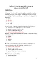
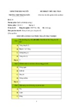
3.4.1. Knee diameters
Knee diameters increase (mm)
13
1.5
1
0.5
0
Sau 3
D0
ngày
Sau 5
D5
ngày
Chứng
sinh
học
Negative
control
TD0015 1,2g/kg
Sau 1
tuần
D7
Sau 2
tuần
D14
Sau 3
tuần
D21
Positive
Mô hìnhcontrol
Sau 4
D28
tuần
Sau 5
D35
tuần
Sau 6
D42
tuần
Diclofenac
TD0015 3,6g/kg
Chart 3.1. Change in knee diameters over time
3.4.2. The analgesic, improving knee joint movement effects
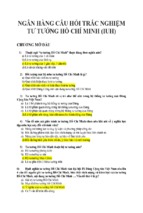
3.4.2.1. The analgesic effect using Randall Selitto method
Pain force (g)
500
400
300
200
100
Trước Sau 1 tuần Sau
3 tuần Sau
4 tuần SauD35
5 tuần Sau D42
6 tuần
D21
D28
D142 tuần Sau
D0
D7
nghiên cứu
Negative
control
Chứng
sinh
học
TD0015 1,2g/kg
Mô
hình control
Positive
TD0015 3,6g/kg
Diclofenac
Chart 3.2. Change in force to inflict pain over time
3.4.2.2. The effect on improving knee joint movement using Dynamic Plantar
Aesthesiometer
In group 1, time and force to inflict pain causing the rats to lift their feet
from the Von-Frey filament displayed no difference at all timepoints
compared to D0. In group 2, after 5 weeks and 6 weeks of MIA injection, the
reaction time to pain and force to inflict pain was significantly increased due
to the damaged degenerative knee joints (p <0.01). In TD0015 at 2 doses of
1,2g/kg and 3,6g/kg, 6 weeks after MIA injection, the reaction time to pain
14
Time to jump (s)
and force to cause pain decreased significantly compared to animals in group
2 (p <0.05). This effect was equivalent to diclofenac 3mg/kg.
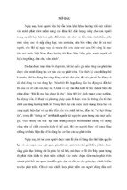
3.4.2.3. The effect of improving knee joint movement using Hot plate
The number of failed jumps in group 2 increased over time compared to
group 1, especially after MIA injection from D21 to D42. Diclofenac 3mg/kg
and TD0015 at 2 doses tended to reduce the number of failed jumps,
especially at week 3, week 5 and week 6 after MIA injection. TD0015 at the
dose of 3,6g/kg displayed more effects than the dose of 1,2g/kg.
10
9
8
7
6
5
4
3
2
1
0
D0
D42
Trước tiêm SauD7
1 tuần SauD14
2 tuần Sau D21
3 tuần Sau 4D28
tuần Sau 5D35
tuần Sau 6 tuần
MIA
Negative control
MôPositive
hình control
Diclofenac 3mg/kg
Chứng sinh học
TD0015 1,2g/kg
TD0015 3,6g/kg
Chart 3.3. Time for the rats to jump off the hot plate
Table 3.5. The height achieved when the rats jump from the hot plate
Group
(n = 10)
Neg.
control
Pos.
control
Diclofenac
3mg/kg
TD0015
1,2g/kg
TD0015
3,6g/kg
D0
22,20 ±4,34
23,50 ±5,80 22,50 ±4,20 23,70 ±4,45
23,50 ±3,63
D7
21,40 ±5,83
19,20 ±4,16 22,30 ±4,99 19,80 ±4,05
21,85 ±4,82
D14
22,40 ±5,32
21,50 ±5,10 20,70 ±4,45 19,40 ±3,92
20,10 ±5,17
Height D21
(cm) D28
20,80 ±3,74
19,70 ±5,74 20,40 ±2,63 19,90 ±2,18
19,80 ±2,82
D35
21,60 ±2,46
19,30 ±3,43 20,90 ±4,20 19,10 ±2,33 19,80 ±1,81
21,10 ±2,92
23,30 ±5,10 18,50 ±2,01*
20,40 ±3,34 21,70 ±4,22 ∆
∆
17,00 ±
20,10 ±3,45
18,40 ±3,44 19,30 ±2,06 ∆
∆
2,36**
*,**,∆: p < 0,05, p < 0,01 compared to neg.control, pos.control (Student’ t-test)
D42
22,10 ±3,51
3.4.3. Effect on cytokine levels
6 weeks after MIA injection, diclofenac 3mg/kg and TD0015 at the
15
dose of 3,6g/kg reduced IL-1β and TNF-α levels significantly compared
to group 2 (p <0.05). In rats taking TD0015 at the dose of 1,2g/kg, IL1β and TNF-α levels tended to decrease compared to animals in group 2
but the difference was not statistically significant (p> 0.05).
*
Cytokines (pg/ml)
250
∆
200
∆
150
IL-1β
100
TNF-α
50
*
∆
∆
0
Mô hình Diclofenac TD0015
Chứng Pos.control
Neg.control
sinh học
1,2g/kg
TD0015
3,6g/kg
Chart 3.4. Cytokine levels
3.4.4. Histopathology of knee joints
3.4.4.1. 2 weeks after MIA injection
In group 1, the level of lesions was minimal. In group 2, the
subchondral lesions, proteoglycan damage, the formation of osteophytes,
and chondrocyte damage were mild to moderate. This difference was
statistically significant compared to animals in group 1 (p <0.05, p <0.01,
p <0.001). The synovial membrane inflammation in group 2 was worse
than group 1 but the difference was not statistically significant (p> 0.05).
Negative control
Positive control
Figure 3.1. Histopathology of knee joints 2 weeks after MIA injection
(HE x 20)
16
3.4.4.2. 6 weeks after MIA injection
Negative control
TD0015 1,2g/kg
Positive control
Diclofenac 3mg/kg
TD0015 3,6g/kg
Figure 3.2. Histopathology of knee joints 6 weeks after MIA injection (HE x 20)
In group 1, the level of lesions is minimal. In group 2, the lesions
were from mild to moderate, significantly higher than group 1 (p
<0.001). In group 3 (diclofenac 3mg/kg), the degree of damage
decreased compared to group 2 (p <0.05). In the group taking TD0015
1,2g/kg, the damage level decreased compared to group 2, especially
proteoglycan structure, cartilage cells and synovial membrane (p
<0.01). In the group taking TD0015 3,6g/kg, all indicators decreased
markedly compared to group 2 (p <0.01), this effect was stronger than
diclofenac 3mg / kg and TD0015 1,2g/kg
Chapter 4. DISCUSSION
4.1. Acute and subchronic toxicity of TD0015 in animals
4.1.1. Acute toxicity
The toxicity of TD0015 may be caused by the toxicity of each
medicinal herb or the interaction of medicinal herbs in this formulation.
Several studies worldwide have shown that most of the herbs in TD0015
have a high LD50, even with a parenteral route. The combination of
herbs in TD0015 did not show any acute toxicity on Swiss mice at the
given doses in this current study. It may be due to the low amount of each
herb in the product. Mice were administered TD0015 from 15g/kg/day to
17
37,5g/kg/day (the largest dose available for mice to take with a gavage
feeding needle), but no mice died and no signs of toxicity were found.
The LD50 value of TD0015 was estimated> 37,5 g/kg body weight. The
recommended human dose is 10 g/day, equivalent to 2,4 g/kg/day in
mice. Thus, white mice ingested up to 15,625 times the dose used in
humans but there was no expression of acute toxicity.
4.1.2. Subchronic toxicity
4.1.2.1. Body weight and clinical observation
After 60 days, rats taking TD0015 at 2 doses reduced food
consumption compared to group 1 but did not show any abnormalities.
The decrease in food intake led to a decrease in the weight gain of the
corresponding rats, but the weight remained stable, not decreased
compared to D0. Among the medicinal herbs included in TD0015, the
Radix Codonopsis pilosulae and Ramulus Cinnnamomi have been
reported to reduce rats' weight gain but the mechanism has not been
clarified. Although not gaining weight compared to group 1, TD0015 at
doses of 1,2 g /kg/day and 3,6 g /kg/day did not adversely affect the
overall condition of rats in 90 days of treatment.
4.1.2.2. Effects on hematopoietic function
In this study, we conducted quantitative blood components including
red blood cell count, hemoglobin content, hematocrit, white blood cell
count, the proportion of lymphocyte and neutrophils, and platelet count.
TD0015 at doses of 1,2g / kg/day and 3,6g / kg/day orally continuously
for 90 days did not change peripheral blood indices in the hematological
tests, displayed no harms to the hematopoietic system.
4.1.2.3. Effects on liver function and liver damage
One of the methods for assessing the degree of hepatocyte damage
is to quantify the concentration of liver-derived enzymes in the serum
(AST, ALT). Besides, the liver is an organ with many functions relating
to the metabolism of substances, including protein and lipid
metabolism. The liver also participates in the synthesis and secretion of
bile. The excretion capacity of the liver was assessed by quantifying
serum bilirubin levels. TD0015 at doses of 1,2g/kg and 3,6g/kg
administered for 90 consecutive days did not reduce albumin
concentration, did not affect bilirubin concentration and AST, ALT
activity. Serum total cholesterol levels of both treatment groups after 90
days of taking the drug were significantly lower than D0 and compared
18
with group 1 at the same timepoint (p <0,05), but still within normal
limits of rats. A number of studies have demonstrated the cholesterollowering effects of the medicinal ingredients in TD0015 such as
paeoniflorin in Radix Paeoniae lactiflorae; saponins, polysaccharides in
Radix Achyranthis bidentatae; hesperidin in PericarpiumCitri
reticulatae. No changes in the overall structure of the heart, lungs, liver,
spleen, pancreas, kidneys and the digestive system were observed in
mice of all 3 groups. The rat livers had no microscopic lesions.
4.1.2.4. Effects on kidney fuction
Evaluation of renal function when taking any drugs is required,
often using quantitative blood creatinine assay. TD0015 of both doses
did not change creatinine levels after 90 days of treatment (p> 0,05),
without adversely affecting the glomerular filtration function.
Histopathology of the kidney was within normal limits, showing that
TD0015 did not affect the function and structure of the rat kidney.
From the above research results, TD0015 at doses of 1,2g / kg/day
and 3,6g/kg/day taken continuously for 90 days did not cause
subchronic toxicity in rats. Thus, TD0015 can be classified as non-toxic
drug for long-term use (90 days). This conclusion is consistent with
some studies in the world regarding the long-term toxicity of each
medicinal herb in TD0015.
4.2. The anti-inflammatory effect of TD0015
4.2.1. The acute anti-inflammatory effect
* Carrageenin-induced rat paw oedema model
Aspirin 200mg/kg demonstrated an acute anti-inflammatory effect
that appeared early but did not last long. TD0015 at the dose of 1,2 g /kg
reduced paw edema after 4 hours but the effect was the most apparent at
24 hours after the injection, with the level of edema suppression up to
40,7%. TD0015 at the dose of 3,6 g/kg displayed this effect sooner than
the lower dose; 2 hours after the injection, it reached 24,33% inhibition of
the oedema, inferior to aspirin. The inhibition increased to 40.3% after 24
hours, which was equivalent to TD0015 at 1,2g/kg. In other words,
TD0015 3,6g/kg had a better acute anti-inflammatory effect with an early
and longer-lasting result. TD0015 at the clinical dose of 1,2g/kg
demonstrated a slower anti-inflammatory effect.
19
* Carrageenin-induced peritonitis model
TD0015 at both doses and aspirin 200mg/kg showed the effect of
reducing the volume of inflammatory exudate, the number of white
blood cells and the quantity of protein compared to the control group (p
<0,01). Thus, both doses of TD0015 had a superior anti-inflammatory
effect on the model of peritonitis, with equivalent effectiveness. The
anti-inflammatory mechanism of TD0015 may be due to its ability to
decrease vascular permeability, inflammatory exudate, and the amount
of protein in the exudate. It also inhibits the migration of white blood
cells into inflamed tissues through limiting the production of chemical
intermediates such as NO, PG E2, IL8, IL-12 or histamine, and 5hydroxytryptamine, which can be the effects of Cortex Phellodendri
amurensi, Radix Paeoniae lactiflorae, Radix Gentianae macrophyllae.
4.2.2. The chronic anti-inflammatory effect
Drugs with chronic anti-inflammatory effects were reported to
inhibit the formation of granulomas and reduce the mass of granulomas
in contrast to that of the control group. The study results showed that in
mice taking TD0015 at doses of 2,4g / kg/day and 7,2g /kg/day, the
weights of granulomas were reduced both before and after drying. The
effect of TD0015 at 7,2g/kg was stronger than that of 2,4g/kg, and was
equivalent to methylprednisolone 10mg / kg.
The results obtained in this study were consistent with other
research on chronic anti-inflammatory effects of each medicinal herb in
TD0015 such as Cortex Phellodendri amurensi, Radix Paeoniae
lactiflorae, Radix Gentianae macrophyllae.
4.3. The analgesic effect of TD0015
4.3.1. The analgesic effect of TD0015 using Hot plate
The hot plate model used heat as a pain agent to evaluate the central
analgesic effect of the drug. TD0015 at two doses of 2,4g /kg /day and
7,2g /kg/day tended to reduce pain, however the effects were not clear.
Therefore, according to this model, TD0015 did not show analgesic
effects through thermal action on the skin. Medicinal herbs in TD0015
may need a longer exposure to be effective. TD0015 was administered
for 5 days, which may not be enough to express the analgesic effect.
4.3.2. The analgesic effect of TD0015 using Von-Frey filament
This method assessed the central analgesic effect of TD0015 by
using a mechanical agent to stimulate pain. The effect was evaluated
20
through the time response to pain and the pain intensity in mice. TD0015
at doses of 2,4g /kg/day and 7,2g /kg/day significantly increased the force
to inflict pain and pain response time in mice causing the pain threshold
meter. This effect was equivalent between the two doses and to codeine
20mg/kg. Thus, on this model, TD0015 had a central analgesic effect
with the model of mechanical pain.
4.3.3. The analgesic effect of TD0015 using acetic acid
The model of pain induced by acetic acid was used to evaluate the
peripheral analgesic effect, using acetic acid to stimulate local
inflammation and pain. Pain was caused by chemicals that can stimulate
macrophages and mast cells to migrate to the peritoneum and release
pain-causing substances such as TNF-α, IL-1β, and IL-8. TD0015 at
doses of 2,4g/kg/day and 7,2g/kg/day reduced the number of painful
cramps at all study points.
From the above results, TD0015 expressed the analgesic effect by
both central and peripheral mechanisms. Some medicinal herbs have
been shown to have analgesic effects such as Cortex Phellodendri
amurensi, Radix Paeoniae lactiflorae…
4.4. The anti-osteoarthritis effect of TD0015
4.4.1. MIA-induced knee osteoarthritis model
In this thesis, the MIA-induced knee OA model was conducted for
the first time in Vietnam, using MIA - a metabolic inhibitor injected into
the knee joints of rats. MIA inhibits the activity of glyceraldehyde-3phosphate dehydrogenase in articular cartilage, causing cartillage and
subchondral bone damage. Many studies have shown that MIA
3mg/single injection is the most effective in creating the OA model.
The diagnosis of OA in animals is mainly based on the following
factors: Assessing symptoms of inflammation, pain, limited movement;
Quantifying specific cytokine indicators in osteoarthritis; Image
exploration: ultrasound, X-ray; Assessing anatomical lesion of the joints
is a gold standard on an experimental model of osteoarthritis. In this
thesis, we evaluated criteria on pain, inflammation, movement restriction,
some specific cytokines, and knee joint histopathology.
Inflammatory indices in the knee osteoarthritis model were
assessed using the diameter of the knee joints in rats. The results
showed that knee joints in group 2 increased significantly compared to
that of group 1, the peak time was 5 days after MIA injection, consistent
- Xem thêm -