The ubiquitin-proteasome pathway of protein degradation is essential for eukaryotes and involves cellular
machinery as elaborate as that required by protein synthesis. Critical molecular steps, however, are
emerging from the discovery of mutations that cause problems ranging from cancer to hypertension to fat
facets1. On page 47 of this issue, Kazumasa Saigoh and colleagues2 broaden the spectrum by discovering
that a mutation in the ubiquitin carboxy-terminal hydrolase gene (Uchl1) causes gracile axonal dystrophy
(gad) in mice.
The gad mouse is a simple genetic model with features reminiscent of inherited neurodegenerative disease
in human3. It develops subtle sensory ataxia, progressing to motor ataxia. Later, axon terminals of the
dorsal root ganglia degenerate, followed by neurons that comprise the gracile nucleus of the medulla
oblongata region4. The brain of the gad mutant contains 'spheroid bodies' and dots of ubiquitin-conjugated
protein in 'dying' terminals and axons5, reminiscent of the often beautiful, varied dots and swirls of
abnormal ubiquitin-conjugated protein found in the post-mortem brains of people with inherited
neurodegenerative diseases. The dots seem to hold a clue to the pathogenic process 1, 6, 7, 8, but the
discovery of unrelated genes which, when mutated, cause more than a dozen of these disorders has
failed, in each instance, to directly link disease mechanism to defects in the cellular machinery that
degrades proteins.
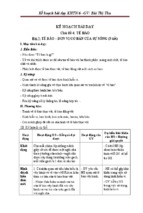
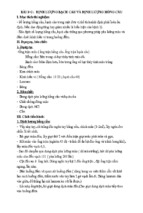
The ubiquitin-proteasome system. A stepwise model of the ubiquitin-proteasome pathway 1 depicting (step 1) release of ubiquitin from
pro-ubiquitin during biosynthesis (steps 2−5) conjugation of ubiquitin to (a) substrate(s) and (steps 6,7) degradation of the
polyubiquitinated-substrate by the 26S proteasome complex (19S, 20S, 19S). Conjugation involves activation of ubiquitin by ubiquitinactivating enzyme E1 (step 2), transfer of activated-ubiquitin to a specific E2 ubiquitin-conjugating enzyme (UBC; step 3), transfer of
activated ubiquitin from E2 to a substrate-specific E3 ubiquitin-ligase (step 4) and formation of a substrate-E3 complex and biosynthesis of
the polyubiquitin chain (step 5). Degradation includes binding of the polyubiquitinated-substrate to the 19S complex (step 6), proteolysis to
short peptides by the 20S complex, and release of recycled ubiquitin (step 7) from 'end' proteolytic products
The identity of the gad gene defect, therefore, comes as a welcome surprise. Saigoh et al. homed in on
the gad locus on chromosome 5 by following co-inheritance of the gad phenotype with polymorphic DNA
markers in a classic genetic linkage-based cloning strategy. Their reward is an in-frame deletion in Uchl1
that results in a truncated form of ubiquitin hydrolase, a thiol protease that participates in ubiquitin
biosythesis and in protein degradation.
This discovery raises new issues. As usual, the molecular consequence of the mutation as mode of action
needs to be delineated. A simple loss of UCH-L1 activity is the most parsimonious explanation. But, as the
authors point out, mRNA is synthesized from the mutant Uchl1 allele at normal levels, which makes it hard
to reject a mechanism involving the predicted truncated UCH-L1 product. The larger issue, which remains
a key puzzle for cloned genes whose mutation causes most neurodegenerative disease, is to discover why
the gad genetic defect affects neurons specifically. This is not an easy challenge. UCH-L1's broad
expression pattern in brain and testis differs from other UCH isozymes, hinting at unrecognized,
specialized activities in neurons. At the same time, its expression in many brain regions does not help one
to understand why its mutation affects the gracile nucleus region.
If we assume the simple loss of UCH-L1's hydrolase activity, a picture of how aberrant Uchl1 may affect
cellular metabolism can be envisaged in the context of the 'entire' ubiquitin-proteasome system. This
complex pathway can be considered to be a series of steps, comprising a cycle of polyubiquitination and
de-ubiquitination, that entails ubiquitination and degradation of a specific substrate (see figure). In this
model1, UCH-L1 activity is required at two crucial steps. Initially, the enzyme co-translationally releases
fused ubiquitin from pro-ubiquitin chains formed during ubiquitin biosythesis. At a later point, UCH-L1
frees ubiquitin from degraded end products containing small molecules (amines and thiols). Loss of UCHL1 hydrolase activity seems likely to produce quantitative and qualitative changes in ubiquitin 'pools' that
fuel the ubiquitin-proteasome system. How these alterations provoke selective gad pathology is less
evident because the pathway should also be altered in brain cells that remain unaffected. Perhaps, as
Saigoh and colleagues surmise, a deficit in the degradation of a specific substrate or, alternatively,
accumulation of an improperly 'de-ubiquitinated' product will explain the toxic effect on select neurons.
Numerous scenarios can be imagined if future studies reveal additional, specialized activities for UCH-L1 in
neurons that may affect pathways other than protein degradation.
But what of the abnormal ubiquitin-conjugated/proteasome dots in gad brain and in human
neurodegenerative diseases that hint of death by failure to degrade proteins? As the authors point out,
loss of UCH-L1 activity provides a direct explanation for the formation of deposits in the gad mouse. In
contrast, with Parkinson disease, Alzheimer disease, amyotrophic lateral sclerosis, and at least seven
'polyglutamine' diseases, including Huntington disease, the emerging connection is indirect. A mutationinduced structural change appears to change the protein's aggregation-properties and/or processing, with
consequences for degradation1, 6, 7, 8.
Connecting the 'dots' to the pathogenic process—for gad and the other diseases—is trickier. The mutationinduced property that propels their formation may be the culprit, but the dots themselves may be either a
cause or a consequence of the disease process1, 6, 7, 8. For example, with respect to Huntington disease, the
glutamine-induced structural property that promotes aggregation of huntingtin's amino terminus is
dependent on glutamine threshold, length-dependence and dosage, identical to those of the disease
mechanism. In this case, the property that propels aggregate-formation seems critical to pathogenesis,
whether the aggregates themselves are toxic or not9.
The key to gad, and all of the neurodegenerative diseases, is neuronal specificity. In each disease,
mutations in widely expressed gene products target discrete neurons, making a general, 'boiler-plate'
model of neurodegenerative disease unlikely. Impaired protein degradation produces the compelling
protein-aggregates, but the 'dots' may be downstream of the critical early events that culminate in
selective neuronal cell death. Nevertheless, the finding that the gad defect is a mutation in Uchl1 promises
to reveal new neuron-specific constituents of the ubiquitin-proteasome system that may yet connect 'dots'
to pathogenesis. Certainly, the discovery bodes well for our understanding of this complex and essential
cellular pathway in the nervous system.
REFERENCES
1. Ciechanover, A. EMBO J. 17, 7151−7160 (1998). | Article | PubMed | ChemPort | Add to Connotea
(beta) |
2. Saigoh, K. et al. Nature Genet. 23, 47−51 (1999). | Article | PubMed | ISI | ChemPort | Add to
Connotea (beta) |
3. Yamazaki, K. et al. Proc. Soc. Exp. Biol. Med. 187, 209−215 (1988). | PubMed
| ISI | ChemPort | Add to Connotea (beta) |
4. Miura, H. et al. Neuropath. Appl. Neurobiol. 19, 41−51 (1993). | ISI | ChemPort |
5. Wu, J. et al. Alzheimer's Res. 2, 163−168 (1996).
6. Alves-Rodrigues, A. et al. Trends Neurosci. 21, 516−520 (1998). | Article | PubMed
| ISI | ChemPort | Add to Connotea (beta) |
7. Ross, C.A. Neuron 19, 1147−1150 (1997). | Article | PubMed | ISI | ChemPort | Add to Connotea
(beta) |
8. Sisodia, S.S. Cell 95, 1−4 (1998). | Article | PubMed | ISI | ChemPort | Add to Connotea (beta) |
9. Huang, C.C. et al. Som. Cell Mol. Genet. 24, 217−233 (1998). | Article | PubMed
| ISI | ChemPort | Add to Connotea (beta) |
Trypsin Activation Peptide (TAP) in Acute Pancreatitis: From
Pathophysiology to Clinical Usefulness
Jean Louis Frossard
Division of Gastroenterology, Geneva University Hospital. Geneva, Switzerland
Acute pancreatitis is a common digestive disease which is usually diagnosed when there is acute abdominal
pain associated with a concomitant rise of serum amylase and lipase levels [1, 2]. However, up to 20% of
patients with acute pancreatitis may have normal serum enzyme concentrations [3]. After exposure to a trigger
event (mainly alcohol and gallstone migration into the common bile duct), injury to the gland occurs extremely
rapidly and is usually complete at the time of admission. For the past 10 years, research aimed at understanding
the early events which initiate acute pancreatitis has provided new information which has led to the recent
development of potentially useful diagnostic tools. In the mid 1990s, the urinary concentration of trypsinogen
and trypsinogen activation peptide (TAP) was shown to be more sensitive and specific in diagnosing acute
pancreatitis than serum amylase and lipase concentrations [4, 5, 6]. Since then, urinary trypsinogen and urinary
TAP represent good alternative tools for clinicians in this situation, but the detection kits are expensive and not
available in every hospital.
Acute pancreatitis is also a disease of variable severity, while approximately 80% of patients experience mild
attacks which resolve themselves with little morbidity, the remaining 20% [7] suffer from severe disease with
mortality rates as high as 30% [8]. Early prediction of the severity of an attack of acute pancreatitis remains the
main goal for clinicians in charge of such patients. The complexity of using multifactorial scales, including
Ranson [9], Glasgow [10] and APACHE II [11] scoring systems, and the fact that CT scanning is expensive,
exposes the patient to ionizing radiation and lacks sensitivity and specificity in the early stage of the disease
[12], account for the increasing interest shown in serum markers to predict the severity of an attack. If severe
attacks were detected at an early stage, aggressive and efficient measures could be implemented without undue
delay. Thus, such patients will probably benefit from admission to tertiary center, prophylactic antibiotic
administration [13], early enteral nutrition [14] and early endoscopic retrograde cholangiopancreatography in
pancreatitis of suspected biliary origin [15]. In the recent study of Neoptolemos et al. [16], urinary TAP
concentration measurement is proposed as a valuable predictive factor inasmuch as it provided accurate severity
prediction in 172 patients with acute pancreatitis (35 with a severe form) 24 hours after the onset of an attack
(70% accuracy at 24 hours).
In this article, we would like to briefly review the pathophysiology of acute pancreatitis and try to determine the
effectiveness in using TAP either as a diagnostic tool or prognostic indicator in acute pancreatitis.
TAP: An Indicator of the First Molecular Event during Experimental Pancreatitis ?
Trypsinogens are pancreatic proteases that can initiate the autodigestive cascade characterizing acute
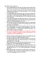
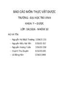
pancreatitis. TAP corresponds to the N-terminal region of the peptide released by the activation of trypsinogen
into active trypsin (Figure 1). Normally, this 7-10 amino peptide is released only when trypsinogen has reached
the small intestine where it is activated by the brush-border enzyme enterokinase. This small cleavage molecule
is immunologically completely distinct from the same sequence within trypsinogen allowing for detection of
TAP in situ. In acute experimental pancreatitis, recent reports have shown that one of the first steps during acute
pancreatitis consisted of inappropriate and premature activation of trypsinogen into active trypsin within the
pancreas resulting in the release of TAP into the peritoneum, plasma and urine [17, 18, 19].
Figure 1. Trypsinogen could be either activated into active trypsin either by the brush-border enzyme enterokinase in the small
intestine or by cathepsin B, a lysosomal enzyme present in acinar cells. Another mechanism of trypsinogen activation, which is a
unique feature of human trypsinogen, consists of trypsinogen autoactivation. This finding may be more relevant to human pancreatitis
whereas cathepsin B mediated trypsinogen activation is more relevant to rodent models of pancreatitis. Once trypsin is activated, it
can catalyze the activation of other digestive pro-enzymes as well as trypsinogen itself, initiating the auto-digestion of the gland.
Recent reports claim that the colocalization of trypsinogen and cathepsin B in the same compartment could result in premature
activation of trypsinogen and leads to acute pancreatitis.
Research directed at understanding the early molecular mechanisms which drive acute pancreatitis from the
trigger event to the phase in which it manifests itself is the subject of controversy. There are two major theories
which have been postulated as to the site and mechanism of trypsinogen activation: the co-localization theory
which may be of relevance only in rodents [17, 20] and the trypsinogen autoactivation [21, 22], a unique feature
of human trypsinogen, which may be more relevant to human pancreatitis.
The co-localization theory claims that intraacinar cell activation of digestive enzymes is initiated by lysosomal
hydrolases acting on trypsinogen either after fusion of the zymogen granules and lysosomes or because
lysosomal enzymes are not segregated from the secretory pathway with complete fidelity (missorting
mechanism) [17, 18, 19, 23, 24]. Using very dissimilar models of pancreatitis, co-localization of digestive
enzymes with the lysosomal enzyme cathepsin B was found to be an early phenomenon preceding cell injury in
rodents [24]. This theory is based on the following findings: 1) adjunction of cathepsin B, a lysosomal enzyme,
is capable of activating digestive enzymes from dogs [25] or human pancreatic extracts [26]; 2) cell
fractionation experiments show co-localization of these different enzymes in the same sedimentation fraction
[17, 27]; 3) inhibition of either trypsin or cathepsin B can effectively prevent trypsinogen activation [24]; 4) this
latter speculation is also supported by recent observations that cathepsin B knockout mice are partially protected
against cerulein-induced pancreatitis because cerulein-induced cathepsin B-mediated activation of trypsinogen
cannot occur in these animals [28]. Taken together, these observations suggest that the initiation of acute
pancreatitis occurs in a compartment containing both of these enzymes.
The second theory postulates that trypsinogen activation occurs in the normal pathway under low pH conditions
and becomes pathological only with a secretory blockade. Under normal conditions, a fraction of the human
trypsinogen autoactivates to active trypsin. Trypsin can catalyze a cascade of trypsinogen activation as well as
activate all other proenzymes leading to the autodigestion of the gland. This process is regulated by at least two
different lines of defense. The first one is pancreatic secretory trypsin inhibitor (PSTI) which is now referred to
as SPINK1 (serine protease inhibitor, Kazal type 1) [29]. When levels of trypsin activity are low, SPINK1
inhibits trypsin and prevents further autoactivation of trypsin and other proenzymes within the pancreas. During
excessive trypsinogen activation, the SPINK1 inhibitory capacity is overwhelmed and trypsin activity keeps
increasing. The second line of defense is represented by trypsin itself. Indeed, to prevent uncontrolled enzyme
activation, trypsin and trypsin-like enzymes, by means of a feedback mechanism, hydrolyze the chain
connecting the two globular domains of the trypsin at R122H. This results in permanent inactivation of trypsin
and prevents subsequent activation of other proenzymes. Recent reports by Whitcomb et al. [23] have strongly
suggested that premature trypsin activation also plays a pivotal role in human acute pancreatitis. This group has
identified two trypsinogen mutations that result in inactivation-resistant trypsin in patients with hereditary
pancreatitis [23]. During excessive trypsinogen activation, the R122H trypsin recognition site is mutated and,
therefore, the trypsin cannot be inactivated leading to autodigestion of the gland and pancreatitis. Furthermore,
although SPINK1 mutations are as high as 2% in the general population, they are clearly associated with
familial and chronic pancreatitis [30]. The last paper by Whitcomb’s group [30] suggests that SPINK1
mutations are disease modifying, possibly by lowering the threshold for pancreatitis from other genetic or
environmental factors, but, by themselves, they do not cause disease.
Taken together, all these observations suggest that one of the earliest events during acute pancreatitis consists of
inappropriate and premature activation of trypsinogen into active trypsin within the pancreas resulting in the
release of TAP into the peritoneum, plasma and urine [17, 18, 19]. Thus, plasma TAP concentration seems to be
among the best and earliest markers of acute pancreatitis. In this setting, it is reasonable to consider TAP as a
sensitive and specific diagnostic tool of an attack of pancreatitis. However, because TAP is a 7-10 amino-acid
peptide, one needs to keep in mind that it is rapidly excreted in urine and its value is therefore limited to the first
24-48 hours after the onset of the symptoms. Moreover, its detection in plasma is more difficult than in urine.
Pancreatic Products as Diagnostic Tools of an Attack of Acute Pancreatitis
Even if most patients with acute pancreatitis have an uncomplicated outcome, early diagnosis of acute
pancreatitis is important because 20% of patients will develop the severe disease with local or systemic
complications [7]. Therefore, immediate diagnosis of severe pancreatitis should be assessed in order to optimize
therapy and to prevent organ dysfunction.
Although amylase and lipase are important for the diagnosis of acute pancreatitis, these enzymes are imprecise
in certain cases [31]. In a series of 352 consecutive cases of acute pancreatitis confirmed by CT scan, 19% of
the patients had normal amylase concentrations in serum upon admission [3]. Acute pancreatitis with normal
amylasemia is characterized by a high prevalence of alcoholic origin [3]. In the study of Pezzilli et al. [32],
serum amylase and lipase levels were able neither to establish the etiology nor to predict the severity of acute
pancreatitis. Recent studies support the view that proteolytic enzymes have a role in the pathophysiology of
pancreatitis and the concentration of trypsinogen in serum was shown to reflect pancreatic injury [5, 33]. The
accuracy of the urinary trypsinogen-2 dipstick test in differentiating between patients with acute pancreatitis,
acute abdominal disease of extrapancreatic origin or no abdominal disease was assessed by Hedström et al. [34]
with a sensitivity of 91% and a specificity of 95% (Table 1). In a study done by the same group [35] concerning
patients with acute abdominal pain, a negative dipstick test for urinary trypsinogen-2 ruled out acute pancreatitis
with a high degree of probability (sensitivity 95%, negative predictive value 99%) (Table 1).
Table 1. Performance of pancreatic enzymes and pancreatic-related products in the diagnosis of acute pancreatitis.
Marker
Sensitivity
Specificity
PPV
NPV
Author
Amylase
85%
91%
-
-
Kemppainen [35]
Amylase
81-85%
87-89%
-
-
Dominguez-Munoz [57]
Lipase
92-95%
95-97%
-
-
Dominguez-Munoz [57]
Phospholipase
34-57%
75-80%
-
-
Dominguez-Munoz [57]
Pancreatitis-associated protein
45-61%
70-83%
-
-
Dominguez-Munoz [57]
Trypsinogen
91%
95%
-
-
Hedström [34]
Trypsinogen
95%
95%
68%
99%
Kemppainen [35]
PPV: positive predictive value
NPV: negative predictive value
TAP assay in urine was first performed in 1990 on 55 patients with acute pancreatitis [6]. A negative result on
admission which was maintained over the first 12 to 24 hours suggested that these patients would either have no
pancreatitis at all or, if so, a very moderate form. Additionally, in the study by Tenner et al. [36], median urinary
TAP at admission was lower in controls than in patients with acute pancreatitis.
TAP as a Prognostic Factor of an Attack of Acute Pancreatitis
All of the causes of acute pancreatitis result in a similar pattern of disease, but the severity of each cannot be
predicted [37]. Most observers believe that the various causes of pancreatitis converge to the same point which
initiates a cascade of events, the nature and extent of which will determine the outcome. TAP has been chosen
as a potential marker of severity, because trypsinogen activation starts within minutes after exposure to a causal
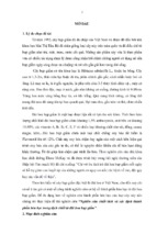
factor. As a result of trypsinogen activation, the trypsinogen and carboxypeptidase B activation peptides
(CAPAP), which are markers of zymogen activation, are released into the serum early in the course of the
disease. (Figure 2).
Figure 2. The intrapancreatic activation of trypsinogen into active trypsin is a process regulated by at least two distinct mechanisms.
1. The PSTI (pancreatic secretory trypsin inhibitor), now referred as to SPINK1 (serine protease inhibitor, Kazal type 1), is
synthesized with trypsinogen in a ratio of 1:5, and can inhibit the activation of trypsinogen by trypsin. 2. Trypsin itself by means of a
feed-back mechanism can inactivate trypsin and trypsin-like enzymes by hydrolyzing the connecting chain between the two globular
domains of the trypsin. Interestingly, in patients with hereditary pancreatitis, trypsin cannot be inactivated because of a mutation
(R122H) in the connecting chain making its hydrolysis impossible.
Although TAP utility has been reported in three major papers [6, 16, 36], CAPAP represents another activation
peptide that is undergoing evaluation. Indeed, in the last study by Pezzilli et al. [38], the overall sensitivity and
specificity of CAPAP in assessing the severity of an attack of acute pancreatitis were 84.6% and 59.4%
respectively (Table 2).
Table 2. Performance
Marker
of pancreatic enzymes and pancreatic-related products in the prediction of the outcome of
an attack.
Sensitivity Specificity
PPV
NPV
Author
CAPAP
85%
59%
-
-
Pezzilli [38]
TAP
80%
90%
67%
-
Gudgeon [6]
CRP
53%
55%
-
-
Gudgeon [6]
TAP
100%
85%
60%
100%
Tenner [36]
TAP at 24 h
58%
73%
39%
86%
Neoptolemos [16]
CRP at 24 h
0%
90%
0%
75%
Neoptolemos [16]
TAP at 48 h
83%
72%
44%
94%
Neoptolemos [16]
CRP at 48 h
86%
61%
37%
94%
Neoptolemos [16]
TAP + CRP
74%
85%
58%
92%
Neoptolemos [16]
APACHE II
56%
64%
30%
85%
Neoptolemos [16]
IL-6
80%
-
71%
-
Leser [54]
IL-6
70%
-
45%
-
Heath [55]
Polymorphonuclear Elastase
93%
-
80%
-
Dominguez-Munoz [47]
Polymorphonuclear Elastase
71%
-
60%
-
Gross [48]
PPV: positive predictive value
NPV: negative predictive value
The first clinical paper referring to TAP use in human beings was published in 1990 [6]. In that study, urinary
samples were collected within 48 hours after the onset of symptoms. The concentrations of TAP correlated with
subsequent disease severity in 87%. By comparison, C-reactive protein and multifactorial scales at 48 hours
were correct in 55% and 84%. The second study by Tenner et al. [36] showed that the median urinary TAP
within 48 hours after the onset of symptoms was significantly higher in patients with severe pancreatitis than in
patients with mild attacks and control patients. Severe pancreatitis was identified in all patients having a urinary
TAP greater than 10 ng/mL, whereas only 6 of 40 patients with mild pancreatitis had a TAP greater than 10
ng/mL. The authors conclude that urinary TAP is useful in identifying patients with severe acute pancreatitis if
obtained within the first 48 hours following the onset of the symptoms (Table 2). In the third and last study
dealing with TAP, the group of Neoptolemos carried out a multicenter study in 246 patients 172 of whom had
acute pancreatitis (35 severe) and 74 were controls. This study was aimed at comparing urinary TAP to Creactive protein (CRP) and three indices scoring systems, but failed to provide more information than the two
previous papers published in 1990 and 1997 respectively. This study was original in that it gave the
performances of urinary TAP at different time points, including 24 hours after the onset of symptoms. At 24
hours after the onset of symptoms, the sensitivity, specificity, positive predictive and negative predictive values
of the test to show severe acute pancreatitis as compared to mild acute disease were 58%, 73%, 39%, and 86%
for TAP greater than 35 mmol/L, and 0%, 90%, 0%, and 75% for CRP greater than 150 mg/L, respectively. The
results of this study fit well with the concept of Neoptolemos which claims that the preferred characteristics of a
prognostic marker have a high negative predictive value, thus allowing a high proportion of patients with the
mild form of the disease to be followed at home. In clinical practice, the use of a prognostic marker capable of
accurately identifying the patients who will develop severe pancreatitis seems more reasonable and efficient.
The comments by Windsor [39], which appeared as an accompanying commentary of the Neoptolemos paper,
are most welcome. Windsor elegantly demonstrated that comparing likelihood ratios was more appropriate for
identifying the patients who would have a severe outcome than were predictive value or accuracy which are
better suited to population studies. When applied to the Neoptolemos study, the likelihood ratios were all of
similar amplitude and there appeared to be no difference between TAP, CRP and the three scoring systems.
Surprisingly, the likelihood ratio was even better for the combined measurement of TAP and CRP, although
Neoptolemos did not claim this [16] (Table 2). In summary, TAP performed no better than the other methods in
terms of overall accuracy.
Perspectives
Traditional severity scores have been used successfully by most clinicians to predict severe acute pancreatitis.
These scores, which are complicated to use, measure the multiple physiological derangements induced by the
disease. However, to predict the severity of the pancreatic disease itself, before the occurrence of multiple organ
failure, other single factors have been measured. Thus, several biological markers of severity have emerged in
the past 15 years and their ability to provide additional information on the severity of the disease has been
evaluated in numerous clinical studies. Nowadays, CRP [40, 41, 42, 43, 44, 45], neutrophil elastase [46, 47, 48,
49, 50] and interleukin-6 (IL-6) [51, 52, 53, 54, 55] are among the best markers, but they are not immediately
available in most institutions.
Measurement of TAP in acute pancreatitis seems appealing because activation of trypsinogen into active trypsin
has been reported to be among the earliest molecular events leading to acute pancreatitis. It should be noted
that, in experimental pancreatitis, the release of TAP occurs as early as 15 minutes after cerulein administration
in rodents [17]. Although very attractive, TAP measurement does not provide additional information for
predicting the outcome of an attack of pancreatitis when compared with the results obtained using other
markers. Further studies should be performed with larger cohorts of patients in order to determine whether TAP
measurement could usefully replace serum amylase and lipase determinations in assessing the diagnosis and the
prognosis of acute pancreatitis, since TAP is specific to the pancreas and is liberated within a few hours after the
onset of symptoms.
In clinical practice, physicians need a marker able to detect which patient will develop the severe disease [39].
However, in the absence of a clear understanding of the physiopathology of acute pancreatitis [56], other
factors, either pancreatic enzymes, or cyto/chemokines, will emerge in the near future and will also prove useful
in the early prediction of the severity of an attack of acute pancreatitis.
References
1.
2.
Bradley EL III. A clinically based classification system for acute pancreatitis. Arch Surg 1993; 128:586-90. [More details]
Steinberg W, Tenner S. Acute pancreatitis. N Engl J Med 1994; 330:1198-210. [More details]
3.
Clavien PA, Robert J, Meyer P, Borst F, Hauser H, Herrmann F, et al. Acute pancreatitis and normoamylasemia. Not an
uncommon combination. Ann Surg 1989; 210:614-20. [More details]
4.
Hedstrom J, Sainio V, Kemppainen E, Puolakkainen P, Haapiainen R, Kivilaakso E, et al. Urine trypsinogen-2 as marker of
acute pancreatitis. Clin Chem 1996; 42:685-90. [More details]
5.
Hedstrom J, Sainio V, Kemppainen E, Haapiainen R, Kivilaakso E, Schroder T, et al. Serum complex of trypsin 2 and alpha 1
antitrypsin as diagnostic and prognostic marker of acute pancreatitis: clinical study in consecutive patients. Br Med J 1996;
313:333-7. [More details]
6.
Gudgeon AM, Heath DI, Hurley P, Jehanli A, Patel G, Wilson C, et al. Trypsinogen activation peptides assay in the early
prediction of severity of acute pancreatitis. Lancet 1990; 335:4-8. [More details]
7.
Steer ML. Classification and pathogenesis of pancreatitis. Surg Clin North Am 1989; 69:467-80. [More details]
8.
Steinberg WM. Predictors of severity of acute pancreatitis. Gastroenterol Clin North Am 1990; 19:849-61. [More details]
9.
Ranson JH, Rifkind KM, Roses DF, Fink SD, Eng K, Spencer FC. Prognostic signs and the role of operative management in
acute pancreatitis. Surg Gynecol Obstet 1974; 139:69-81. [More details]
10. Blamey SL, Imrie CW, O'Neill J, Gilmour WH, Carter DC. Prognostic factors in acute pancreatitis. Gut 1984; 25:1340-6.
[More details]
11. Wilson C, Heath DI, Imrie CW. Prediction of outcome in acute pancreatitis: a comparative study of APACHE II, clinical
assessment and multiple factor scoring systems. Br J Surg 1990; 77:1260-4. [More details]
12. Ranson JH, Balthazar E, Caccavale R, Cooper M. Computed tomography and the prediction of pancreatic abscess in acute
pancreatitis. Ann Surg 1985; 201:656-65. [More details]
13. Haber PS, Pirola RC, Wilson JS. Clinical update: management of acute pancreatitis. J Gastroenterol Hepatol 1997; 12:18997. [More details]
14. Windsor AC, Kanwar S, Li AG, Barnes E, Guthrie JA, Spark JI, et al. Compared with parenteral nutrition, enteral feeding
attenuates the acute phase response and improves disease severity in acute pancreatitis. Gut 1998; 42:431-5. [More details]
15. Gupta R, Toh SK, Johnson CD. Early ERCP is an essential part of the management of all cases of acute pancreatitis. Ann R
Coll Surg Engl 1999; 81:46-50. [More details]
16. Neoptolemos J, Kemppainen E, Mayer J, Fitzpatrick J, Raraty M, Slavin J, et al. Early prediction of severity in acute
pancreatitis by urinary trypsinogen activation peptide: a multicentre study. Lancet 2000; 355:1955-60. [More details]
17. Hofbauer B, Saluja AK, Lerch MM, Bhagat L, Bhatia M, Lee HS, et al. Intra-acinar cell activation of trypsinogen during
caerulein-induced pancreatitis in rats. Am J Physiol 1998; 275:G352-62. [More details]
18. Krims PE, Pandol SJ. Free cytosolic calcium and secretagogue-stimulated initial pancreatic exocrine secretion. Pancreas
1988; 3:383-90. [More details]
19. Mayer J, Rau B, Schoenberg MH, Beger HG. Mechanism and role of trypsinogen activation in acute pancreatitis.
Hepatogastroenterology 1999; 46:2757-63. [More details]
20. Otani T, Chepilko S, Grendell J, Gorelick F. Codistribution of TAP and the granule membrane protein GRAMP-92 in rat
caerulein-induced pancreatitis. Am J Physiol 1998; 275:G999-1009. [More details]
21. Whitcomb DC. Early trypsinogen activation in acute pancreatitis. Gastroenterology 1999; 116:770-2. [More details]
22. Whitcomb DC. Hereditary pancreatitis: new insights into acute and chronic pancreatitis. Gut 1999; 45:317-22. [More details]
23. Whitcomb DC, Gorry M, Preston R, Furey W, Sossenheimer M, Ulrich C, et al. Hereditary pancreatitis is caused by mutation
in the cationic trypsinogen gene. Nat Genet 1996; 14:141-5. [More details]
24. Saluja AK, Donovan EA, Yamanaka K, Yamaguchi Y, Hofbauer B, Steer ML. Cerulein-induced in vitro activation of
trypsinogen in rat pancreatic acini is mediated by cathepsin B. Gastroenterology 1997; 113:304-10. [More details]
25. Greenbaum L, Hirschkowitz A. Endogenous cathepsin activates trypsinogen in extracts of dog pancreas. Proc Soc Exp Biol
Med 1961; 107:74-6. [More details]
26. Figarella C, Miszczuk-Jamska B, Barrett A. Possible lysosomal activation of pancreatic zymogens: activation of both human
trypsinogens by cathepsin B and spontaneous acid activation of human trypsinogen 1. Biol Chem Hoppe Seyler 1988; 369
(Suppl):293-8. [More details]
27. Saluja A, Hashimoto S, Saluja M, Powers RE, Meldolesi J, Steer ML. Subcellular redistribution of lysosomal enzymes during
caerulein-induced pancreatitis. Am J Physiol 1987; 253:G508-16. [More details]
28. Halangk W, Lerch M, Brandt-Nedelev B, Roth W, Ruthenbuerger M, Reinheckel T et al. Role of cathepsin B in intracellular
trypsinogen activation and the onset of acute pancreatitis. J Clin Invest 2000; 106:773-81. [More details]
29. Witt H, Luck W, Hennies H, Classen M, Kage A, Lass U, et al. Mutations in the gene encoding the serine protease inhibitor,
Kazal type 1 are associated with chronic pancreatitis. Nat Genet 2000; 25:213-6. [More details]
30. Pfutzer R, Barmada M, Brunskill A, Finch R, Hart P, Neoptolemos J, et al. SPINK1/PSTI polymorphisms act as a disease
modifiers in familial and idiopathic chronic pancreatitis. Gastroenterology 2000; 119:615-23. [More details]
31. Clavien PA, Burgan S, Moossa AR. Serum enzymes and other laboratory tests in acute pancreatitis. Br J Surg 1989; 76:123443. [More details]
32. Pezzilli R, Billi P, Miglioli M, Gullo L. Serum amylase and lipase concentrations and lipase/amylase ratio in assessment of
etiology and severity of acute pancreatitis. Dig Dis Sci 1993; 38:1265-9. [More details]
33. Ohlsson K, Eddeland A. Release of proteolytic enzymes in bile-induced pancreatitis in dogs. Gastroenterology 1975; 69:66875. [More details]
34. Hedstrom J, Korvuo A, Kenkimaki P, Tikanoja S, Haapiainen R, Kivilaakso E, et al. Urinary trypsinogen-2 test strip for acute
pancreatitis. Lancet 1996; 347:729-30. [More details]
35. Kemppainen EA, Hedstrom JI, Puolakkainen PA, Sainio VS, Haapiainen RK, Perhoniemi V, et al. Rapid measurement of
urinary trypsinogen-2 as a screening test for acute pancreatitis. N Engl J Med 1997; 336:1788-93. [More details]
36. Tenner S, Fernandez-del Castillo C, Warshaw A, Steinberg W, Hermon-Taylor J, Valenzuela JE, et al. Urinary trypsinogen
activation peptide (TAP) predicts severity in patients with acute pancreatitis. Int J Pancreatol 1997; 21:105-10. [More details]
37. Karne S, Gorelick F. Etiopathogenesis of acute pancreatitis. Surg Clin North Am 1999; 79:699-709. [More details]
38. Pezzilli R, Morselli-Labate AM, Barbieri AR, Platè L. Clinical usefulness of the serum carboxypeptidase B activation peptide
in acute pancreatitis. JOP. J Pancreas (Online) 2000; 1:58-68. [More details]
39. Windsor J. Search for prognostic markers for acute pancreatitis. Lancet 2000; 355:1924-5. [More details]
40. Buchler M, Malfertheiner P, Schoetensack C, Uhl W, Beger HG. Sensitivity of antiproteases, complement factors and Creactive protein in detecting pancreatic necrosis. Results of a prospective clinical study. Int J Pancreatol 1986; 1:227-35.
[More details]
41. De Beaux AC, Goldie AS, Ross JA, Carter DC, Fearon KC. Serum concentrations of inflammatory mediators related to organ
failure in patients with acute pancreatitis. Br J Surg 1996; 83:349-53. [More details]
42. De la Pena J, De las Heras G, Galo Peralta F, Casafont F, Pons Romero F. Prospective study of the prognostic value of C
reactive protein, alpha 1-antitrypsin and alpha 1-acid glycoprotein in acute pancreatitis. Rev Esp Enferm Dig 1991; 79:33740. [More details]
43. Gross V, Leser HG, Heinisch A, Scholmerich J. Inflammatory mediators and cytokines - new aspects of the pathophysiology
and assessment of severity of acute pancreatitis? Hepatogastroenterology 1993; 40:522-30. [More details]
44. Imrie CW. Prognosis of acute pancreatitis. Ann Ital Chir 1995; 66:187-9. [More details]
45. Isenmann R, Buchler M, Uhl W, Malfertheiner P, Martini M, Beger HG. Pancreatic necrosis: an early finding in severe acute
pancreatitis. Pancreas 1993; 8:358-61. [More details]
46. Buchler M, Malfertheiner P, Uhl W, Beger HG. Diagnostic and prognostic value of serum elastase 1 in acute pancreatitis.
Klin Wochenschr 1986; 64:1186-91. [More details]
47. Dominguez-Munoz JE, Carballo F, Garcia MJ, de Diego JM, Rabago L, Simon MA, de la Morena J. Clinical usefulness of
polymorphonuclear elastase in predicting the severity of acute pancreatitis: results of a multicentre study. Br J Surg 1991;
78:1230-4. [More details]
48. Gross V, Scholmerich J, Leser HG, Salm R, Lausen M, Ruckauer K, et al. Granulocyte elastase in assessment of severity of
acute pancreatitis. Comparison with acute-phase proteins C-reactive protein, alpha 1-antitrypsin, and protease inhibitor alpha
2-macroglobulin. Dig Dis Sci 1990; 35:97-105. [More details]
49. Ikei S, Ogawa M, Yamaguchi Y. Blood concentrations of polymorphonuclear leucocyte elastase and interleukin-6 are
indicators for the occurrence of multiple organ failures at the early stage of acute pancreatitis. J Gastroenterol Hepatol 1998;
13:1274-83. [More details]
50. Uhl W, Buchler M, Malfertheiner P, Martini M, Beger HG. PMN-elastase in comparison with CRP, antiproteases, and LDH
as indicators of necrosis in human acute pancreatitis. Pancreas 1991; 6:253-9. [More details]
51. Berney T, Gasche Y, Robert J, Jenny A, Mensi N, Grau G, et al. Serum profiles of interleukin-6, interleukin-8, and
interleukin-10 in patients with severe and mild acute pancreatitis. Pancreas 1999; 18:371-7. [More details]
52. Bertsch T, Aufenanger J. Interleukin-6 and phospholipase A2 isoenzymes during acute pancreatitis. Pancreas 1998; 16:557-8.
[More details]
53. Cromwell O, Hamid Q, Corrigan C, Barkans J, Meng Q, Collins P, et al. Expression and generation of interleukin-8,
interleukin-6 and granulocyte-macrophage colony stimulating factor by bronchial epithelial cells and enhancement by IL-1
beta and tumor necrosis factor-alpha. Immunology 1992; 77:330-7. [More details]
54. Leser HG, Gross V, Scheibenbogen C, Heinisch A, Salm R, Lausen M, et al. Elevation of serum interleukin-6 concentration
precedes acute-phase response and reflects severity in acute pancreatitis. Gastroenterology 1991; 101:782-5. [More details]
55. Heath DI, Cruickshank A, Gudgeon M, Jehanli A, Shenkin A, Imrie CW. Role of interleukin-6 in mediating the acute phase
protein response and potential as an early means of severity assessment in acute pancreatitis. Gut 1993; 34:41-5. [More
details]
56. Frossard JL. Trypsinogen activation peptide in acute pancreatitis. Lancet 2000; 356:766-7. [More details]
57. Dominguez-Munoz JE. Diagnosis of acute pancreatitis : any news or still amylase. In: Buchler MW, Uhl W, Friess H,
Malfertheiner P, eds. Acute Pancreatitis, Novel Concepts in Biology and Therapy. Volume 1. 1 st ed. Oxford: Blackwell
Science. 1999:171-9. [More details]
Peptide bond cleavage by PepA and leucine aminopeptidase
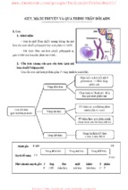
Biological background
E. coli aminopeptidase A (PepA) und bovine lens leucine aminopeptidase (LAP) share ~30% sequence identity.
As the name implies, these exopeptidases cleave the N-terminal amino acids from peptides. Human LAP has
been shown to catalyze postproteasomal trimming of the N-terminus of antigenic peptides for presentation on
MHC class I molecules. Here, interferon-gamma not only promotes proteasomal cleavage but also indices LAP
for N-terminal processing of the peptides.
In addition to the aminopeptidase activity, PepA (but not LAP) has independent DNA-binding functions.
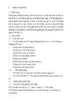
Hexamer structure
E. coli aminopeptidase A (PepA) is a hexameric protein of symmetry 32,
i.e. two-fold molecular axes are perpendicular to a three-fold molecular
axis. In the figure on the left, the view is along the three-fold axis.
The C-terminal domains are shown in blue and the N-terminal domains in
green. A long helix (orange) connects the two domains. The catalytic zinc
ions are shown in yellow.
Compartimentalization
The aminopeptidase active sites (marked by the catalytic zinc ions shown
in red) are located in the center of the hexamer, where a large cavity of 30
Angstrom diameter and 10 Angstrom height is formed. Access to this
cavity is provided by channels. Such a compartimentalization of the
reaction room also occurs in other proteases and in the proteasome (see
review by Larsen and Finley, 1997). In PepA and LAP the
compartimentalization ensures that the enzyme acts only on small
peptides (~6 residues) and not on proteins.
In the left figure a LAP hexamer has been cut open perpendicular to the
threefold axis. The cut protein regions are coloured green and the rest of
the protein surface in blue and white. The active sites are marked in red.
The aminopeptidase active site
The electron density map of LAP at 1.6 Angstrom resolution
reveals many details of the active site. A surprising finding was the
binding of a carbonate ion next to an arginine residue. The
structure of PepA, which was determined later, and kinetic studies
on wild-type PepA and mutant variants showed that this carbonate
ion is not an artifact of the crystallization conditions, but is part of
the active site and it has a functional role.
PepA is activated ~8-fold by physiological concentrations of
bicarbonate ions, i.e. that are present in the cell from dissolved
carbon dioxide.
In the unliganded structure both zinc ions are five-coordinated,
mainly by oxygen atoms from carboxylate side chains, a peptide
carbonyl group and a water molecule. A somewhat unusual metal
ligand is the lysine residues coordinated to Zn2.
Inhibitor binding
Details on the enzyme mechanism were obtained from the
binding of transition-state analogues to the catalytic center.
Shown on the left is the binding mode of leucinal, in which the
carboxylate group of leucine is replaced by an aldehyde group.
The aldehyde group is hydrated to a gem-diol, which mimicks the
gem-diolate group of the transition-state of the reaction.
Both hydroxyl groups of the gem-diol are coordinated to the zinc
ions. In addition, the amino group of the inhibitor is also bound to
one of the zinc ions.
In the transition-state both zinc ions are six-coordinated.
Catalytic mechanism
References
Larsen, C. N. & Finley, D. (1997). Protein translocation channels in the proteasome and other proteases. Cell
91, 431-434.
Sträter, N., Sun, L., Kantrowitz, E. N. & Lipscomb, W. N. (1999). A carbonate ion as a general base in the
mechanism of peptide hydrolysis by dizinc leucine aminopeptidase. Proc. Natl. Acad. Sci. USA 96, 1115111155.
Sträter, N. & Lipscomb, W. N. (1995). Two-metal ion mechanism of bovine lens leucine aminopeptidase: active
site solvent structure and binding mode of L-leucinal, a gem-diolate transition state analogue, by X-ray
crystallography. Biochemistry 34, 14792-14800.
Sträter, N. & Lipscomb, W. N. (1995). Transition state analogue L-leucinephosphonic acid bound to bovine lens
leucine aminopeptidase: X-ray structure at 1.65 Å resolution in a new crystal form. Biochemistry34, 9200-9210
Ribonucleotide reductase (RNR) is an enzyme that controls the cellular concentration of
deoxyribonucleotides. Biosynthesis begins with the building up of essential molecules that RNR processes in a
catalyzed reaction to make deoxyribonucleotides. RNR assembles deoxyribonucleotides for the synthesis of
DNA. The processes are identical in all living organisms. What makes RNR unique from other enzymes is the
need for a free radical.
Deoxyribonucleotides are synthesised on the level of diphosphates. The substrates for RNR are ADP, GDP, CDP
and UDP. dTDP is synthesised by another enzyme (thymidilate synthase) from dUMP.
Structure
The iron-dependent enzyme, ribonucleotide reductase (RNR), is essential for DNA synthesis. Class I RNR
comprises RNR1 and RNR2 subunits, which can associate to form an active heterodimeric tetramer. Since the
enzyme catalyses the de novo synthesis of deoxyribonucleotides (dNTPs), precursors to DNA synthesis, it is
essential for cell proliferation.
Each RNR1 monomer consists of three domains: one mainly helical domain comprising the 220 N-terminal
residues; a second large ten-stranded α/β structure [α-helix and β-sheet, Structural Classification of Proteins
(SCOP)] comprising 480 residues; and a small five-stranded α/β structure comprising 70 residues (Jordan &
Reichard, 1998). RNR2 contains a diferric iron center and a stable tyrosyl radical. In E. coli, the tyrosyl radical
is located at position 122 (Y122) providing the stable radical for the Class I RNR2 subunits (Hogbom et al.,
2001). In A. aegypti, this tyrosyl radical is located at position 184 (Y184) (Pham et al., 2002). The tyrosyl
radical is deeply buried inside the protein in a hydrophobic environment, located close to the iron center that is
used in the stabilization of a tyrosyl radical. The structure of two μ-oxo-linked irons is dominated by ligands
that serve as iron binding sites: four carboxylates [aspartate (D146), glutamate (E177, E240, and E274)] and
two histidines (H180 and H277) (Pham et al., 2002). Association occurs between the C-terminus of RNR2 and
the C-terminus of RNR1 (Jordan & Reichard, 1998). Enzymatic activity is dependent on association of the
RNR1 and RNR2 subunits. The active site consists of the active dithiol groups from the RNR1 as well as the
diferric center and the tyrosyl radical from the RNR2 subunit.
Other residues of RNR2, such as aspartate (D273), tryptophan (W48), and tyrosine (Y356) further stabilize the
active-site tyrosyl radical thus allowing electron transfer (Jordan & Reichard, 1998). These residues help in the
transfer of the radical electron from tyrosine (Y122) of RNR2 to cysteine (C439) of RNR1. The electron
transfer begins on RNR2 tyrosine (Y122) and continues in RNR2 to tryptophan (W48), which is separated from
RNR1 tyrosine (Y731) by 2.5 nanometers. Electron transfer from RNR2 to RNR1 occurs via tyrosine (Y356 to
Y731) and continues on through tyrosine (Y730) to cysteine (C439) in the active site (Chang et al., 2004). Sitedirected mutations of the RNR primary structure indicate that all residues cited above participate in the long
distance transfer of the free radical to the active site (Jordan & Reichard, 1998).
In Aedes aegypti mosquitoes, RNR2 retains most of the crucial amino acid residues, including aspartate (D64)
and valine (V292 or V284), that are necessary in allosteric regulation; proline (P210 and P610), leucine (L453
and L473), and methionine (M603) residues that are located in the hydrophobic active site; cysteine (C225,
C436 and C451) residues that are involved in removal of a hydrogen atom and transfer of the radical electron at
the active site; cysteine (C225 and C436), asparagine (N434), and glutamate (E441) residues that bind the
ribonucleotide substrate; tyrosine (Y723 and Y743) residues that dictate the radical transfer; and cysteine (C838
and C841) residues that are used in the regeneration of dithiol groups in the active site (Pham et al., 2002).
Function
The enzyme ribonucleotide reductase (RNR) catalyzes the de novo synthesis of dNTPs (Nelson & Cox, 2000).
Catalysis of ribonucleoside 5’-diphosphates (NDPs) involves a reduction at the 2’-carbon of ribose 5-phosphate
to form the 2’-deoxy derivative-reduced 2’-deoxyribonucleoside 5’-diphosphates (dNDPs). This reduction is
initiated with the generation of a free radical. Following a single reduction, RNR requires electrons donated
from the dithiol groups of the protein thioredoxin. Regeneration of thioredoxin occurs when nicotinamide
adenine dinucleotide phosphate (NADPH) provides two hydrogen atoms that are used to reduce the disulfide
groups of thioredoxin.
Step 1 = an electron transfer on the RNR2 subunit activates a RNR1 cysteine residue in the active site
with a free radical; Step 2 = the free radical forms a stable radical on C-3, and cysteine residue removes a
hydrogen; Step 3 = a cation is formed on C-2 by transferring a hydrogen from a dithiol group and is
stabilized by the radical, resulting in the loss of H2O from C-2; Step 4 = a hydrogen is transferred from
the dithiol group to reduce the cation C-2; Step 5 = the C-3 radical is reduced by the hydrogen removed
in step 2, and the tyrosyl radical is generated; Step 6 = redoxins transfer two hydrogen to the disulfide
group that restores the original configuration.
Three classes of RNR have similar mechanisms for the reduction of NDPs, but differ in the domain that
generates the free radical, the specific metal in the metalloprotein structure, and the electron donors. All classes
use free-radical chemistry (Jordan & Reichard, 1998). Class I reductases use an iron center with ferrous to ferric
conversion to generate a tyrosyl free radical. Reduction of NDP substrates occurs under aerobic conditions.
Class I reductases are divided into IA and IB due to differences in regulation. Class IA reductases are distributed
in eukaryotes, eubacteria, bacteriophages, and viruses. Class IB reductases are found in eubacteria. Class IB
reductases can also use a radical generated with the stabilization of a binuclear manganese center. Class II
reductases generate a free radical by mechanisms involving 5’-deoxyadenosyl cobalamin (coenzyme B12) and
have a simpler structure than class I and class III reductases. Reduction of NDPs or ribonucleotide 5’triphosphates (NTPs) occurs under either aerobic or anaerobic conditions. Class II reductases are distributed in
archaebacteria, eubacteria, and bacteriophages. Class III reductases use a glycine radical generated with the help
of an S-adenosyl methionine and an iron sulphur center. Reduction of NTPs is limited to anaerobic conditions.
Class III reductases are distributed in archaebacteria, eubacteria, and bacteriophages (Jordan & Reichard, 1998
& Pham et al., 2002). Organisms are not limited to having one class of enzymes. For example, Escherichia coli
have both class I and class III RNR.
Metabolic pathways
Several major pathways lead to the generation of precursors for the de novo synthesis of nucleotides. These
pathways involve the generation of ribose 5-phosphate, carbon dioxide, amino acids and ammonia. Ribose 5phosphate generation begins with a molecule of glucose that is oxidized via the pentose phosphate pathway. The
pentose phosphate pathway produces NADPH for reducing power involved in the catalysis of NTPs to dNTPs,
and to produce ribose 5-phosphate necessary for the synthesis of ribonucleotides. Carbon dioxide is always
available for biosynthesis because its concentration in the blood is kept nearly constant via the bicarbonate
buffer system. An important co-factor for ribonucleotide synthesis is tetrahydrofolate, which is the major
mediator for carbon transfers. Its derivative, folate (a vitamin), cannot be synthesized in mammals. Many forms
of tetrahydrofolate follow pathways that are interconnected. For ribonucleotide synthesis, the N10-formyltetrahydrofolate molecule is necessary for the transfer of formyl groups to the purine ring. Amino groups or
ammonia are donated from the catabolism of amino acids beginning with a dietary protein molecule. The free
ammonia is combined with glutamate by a reaction involving adenosine 5’-triphosphate (ATP) and the activity
of glutamine synthetase, which produces a nontoxic molecule of glutamine that can be transported in the
bloodstream. Glutamine synthetase is present in nearly all organisms and is allosterically regulated by end
products of glutamine metabolism. During synthesis of purines, amino groups are removed from glutamine for
purine rings. Purine ribonucleotides are attached to ribose 5-phosphate during assembly of intermediate
inosinate (IMP) from precursors in the purine pathway, including glutamine, glycine, N10-formyltetrahydrofolate, bicarbonate, aspartate and ATP. Synthesis is catalyzed by large multienzyme complexes.
Purine ribonucleotides are adenosine 5’-monophosphate (AMP) and guanosine 5’-monophosphate (GMP). AMP
is formed from IMP by aspartate donating an amino group (leaving as fumarate) and guanosine 5’-triphosphate
(GTP) providing a phosphate. GMP is formed by the oxidation of IMP at C-2 requiring NAD+. Following
oxidation, glutamine donates an amino group (leaving as glutamate) then ATP provides a phosphate.
Pyrimidine ribonucleotides are formed from an orotate molecule that is assembled from aspartate to form the
pyrimidine ring. Subsequently, orotate is attached to ribose 5-phosphate to yield orotidylate. These two steps are
catalyzed by a large multienzyme complex (CAD). Pyrimidine ribonucleotides are cytidine 5’-monophosphate
(CMP) and uridine 5’-monophosphate (UMP). Orotidylate is decarboxylated to form UMP. UMP and two ATPs
are transferred by kinases to form uridine 5’-triphosphate (UTP). Cytidine 5’-triphosphate (CTP) is formed
from UTP by glutamine donating an amino group (leaving as glutamate) and ATP providing a phosphate. In
some species, ammonia can donate an amino group instead of glutamine. Generation of 2’-deoxythymidine 5’monophosphate (dTMP) occurs by conversion of 2’-deoxyuridine 5’-monophosphate (dUMP). Thymidylate
synthase catalyzes the reaction in which dTMP is formed from dUMP; to provide the carbon atom N5, N10methylene-tetrahydrofolate is oxidized to 7, 8-dihydrofolate. Dihydrofolate reductase (DHFR) is an essential
enzyme that regenerates tetrahydrofolate at the expense of NADPH. Ribonucleoside monophosphates (AMP,
GMP, CMP, and UMP) are phosphorylated to ribonucleoside diphosphates for their particular base by specific
kinases. Ribonucleoside diphosphates are phosphorylated a second time to ribonucleoside triphosphates by
nucleoside diphosphate kinase, which is not specific for their base or for their 2’-carbon of ribose 5-phosphate
and its 2’-deoxy derivative. The activity of nucleoside diphosphate kinase is sequential based on which class of
RNR is used. These metabolic pathways generate the ribonucleotides (ATP, GTP, CTP, and UTP) that are
precursors for dNTPs. Thus, RNR reduces the corresponding NTPs to dNTPs for DNA synthesis. Cellular
concentration of dNTP is much lower then required for DNA replication, and RNR is essential for adequate
precursors during DNA synthesis.
After RNR reduces NDP or NTP the enzyme becomes inactive because a disulfide bond is formed in the active
site. An exchange reaction occurs that reduces the disulfide bond of RNR catalyzed by thioredoxin or
glutaredoxin. RNR gains electrons on the active-site dithiol groups necessary for its activity.
Catalytic Reduction Mechanism
The mechanism that is currently accepted for the reduction of ribonucleotides to deoxyribonucleotides is
depicted in the following scheme. The first step involves the abstraction of the 3’- H of substrate 1 by radical
Cys439. Subsequently, the reaction involves the elimination of one water molecule from carbon C-2’ of the
ribonucleotide, catalyzed by Cys225 and Glu441. In the third step there is a hydrogen atom transfer from
Cys225 to carbon C-2’ of the 2’-ketyl radical 3, after previous proton transfer from Cys462 to Cys225. At the
end of this step, a radical anionic disulfide bridge and the closed-shell ketone intermediate 4 are obtained. This
intermediate has been identified during the conversion of several 2’-substituted substrate analogues, as well as
with the natural substrate (Cerqueira, 2004) interacting with enzyme mutants. The next step is the oxidation of
the anionic disulfide bridge, with concomitant reduction of the substrate, generating 5. The spin density shifts
from the sulphur atoms to the C-3' atom of the substrate, with simultaneous proton transfer from Glu441 to
carbon C-3'. The last step is the reverse of the first step and involves a hydrogen transfer from Cys439 to C-3’,
regenerating the initial radical and resulting in the final product 6. Theoretical models of some steps of these
mechanisms using the full model of the R1 protein can be found at the studies performed by Cerqueira et al.
(Cerqueira, 2005 and Cerqueira, 2006)
Image:RNR Mechanism.jpg
Regulation
Class I RNR comprises RNR1 and RNR2 subunits, which can associate to form a heterodimeric tetramer
(Eklund et al., 1997). RNR1 contains both allosteric sites, mediating regulation of substrate specificity and
activity (Uhlin and Eklund, 1994). Depending on the allosteric configuration, one of the four ribonucleotides
binds to the active site.
Class I RNR is activated by binding ATP or inactivated by binding dATP to the activity site located on the
RNR1 subunit. When the enzyme is activated, substrates are reduced if the corresponding effectors bind
to the allosteric substrate specificity site. A = when dATP or ATP is bound at the allosteric site, the
enzyme accepts UDP and CDP into the catalytic site; B = when dGTP is bound, ADP enters the catalytic
site; C = when dTTP is bound, GDP enters the catalytic site. The substrates (ribonucleotides UDP, CDP,
ADP, and GDP) are converted to dNTPs by a mechanism involving the generation of a free radical.
Regulation of RNR is designed to maintain balanced quantities of dNTPs. Binding of effector molecules either
increases or decreases RNR activity. When ATP binds to the allosteric activity site, it activates RNR. In contrast,
when dATP binds to this site, it deactivates RNR (Jordan & Reichard, 1998). In addition to controlling activity,
the allosteric mechanism also regulates the substrate specificity and ensures the enzyme produces an equal
amount of each dNTP for DNA synthesis (Jordan & Reichard, 1998). In all classes, binding of ATP or dATP to
the allosteric site induces reduction of cytidine 5’-diphosphate (CDP) and uridine 5’-diphosphate (UDP); 2’deoxyguanosine 5’-triphosphate (dGTP) induces reduction of adenosine 5’-diphosphate (ADP); and 2’deoxythymidine 5’-triphosphate (dTTP) induces reduction of guanosine 5’-diphosphate (GDP) (Figure 1).
Interestingly, class IB reductases are not inhibited by dATP because they lack approximately 50 N-terminal
amino acids required for the allosteric activity site (Eliasson et al., 1996). Eukaryotic cells with class IA
reductases have a mechanism of negative control to turn off synthesis of dNTPs as they accumulate. This
mechanism protects the cell from toxic and mutagenic effects that can arise from the overproduction of dNTPs
because changes in balanced dNTP pools lead to DNA damage and cell death (Kunz, 1988 & Meuth, 1989).
Inhibition of RNR1 and RNR2 structure
Generally Class I RNR inhibitors can be divided in three main groups: translation inhibitors, which unable the
formation of the enzyme; dimerization inhibitors that prevent the complexation of the two RNR subunits (R1
- Xem thêm -