Overheads for Section 3
A brief interlude on digestion
Overview of digestion
More digestion facts
Overview of biochemical pathways
Chapter 14
Glycolysis
The glycolytic pathway
Another view of the glycolytic pathway
The test version
Details of one individual reaction step
Enzyme compexes facilitate channeling
The energy landscape of glycolysis
The "metabolic" regulation of glycolysis
Glycolytic addenda
Other carboyhdrates are funneled into the glycolytic path
Use of galactose
The fates of pyruvate
Gluconeogenesis
Gluconeogenesisb
Pyr to PEP
Two other bypasses
Gluconeogenesis and mitochondria
The Pentose pathway
Alternative views of the Pentose pathway
Chapter 15
The role of glycogen phosphorylase
The mechanism of GP
The breakdown of glycogen
Transporting glucose to the blood
Activating glucose for synthesis
The synthesis of glycogen
Regulation of GP
The alloseric regulation of PFK
Coordinate regulation of PFK and FBP
The role of F2,6BP
Pyruvate Kinase is regulated
Coordinate regulation of glycolysis/gluconeogenesis
Hormonal influence on glucose metabolism
Glycogen synthase is regulated by hormones
Synthesis and hydrolysis of F2,6BP
More on PFK2 and FBPase2
Summary of hormone regulation in the liver
Chapter 16
The TCA cycle in metabolism
Some headlines in C-C chemistry
C C bond reactions in biochemistry
Enolate stabilization in carbon-carbon bonds
TPP modifies α-keto acids for decarboxylation
TPP and decarboxylation
Back to Metabolism
The PDC "linking step"
PDC is a geometric complex
Regulation of PDC
Steps of the TCA cycle
The test version
Mech of citrate synthase, more C-C chem
The energy profile of TCA
Regulation of the TCA cycle
Depletion of TCA intermediates
The anaplerotic reactions
Chemistry of pryuvate carboxylase
The aerobic metabolism of glucose
The glyoxylate cycle
The glyoxylate shunt
Linking TCA and glyoxylate shunt
Chapter 17
Lipids from ingestion, storage, or synthesis
Fat uptake
Lipid mobilization
Chylomicrons
Lipids released from adipocytes
Post lipase chemistry
Carnitine mediated transport
Beta-oxidation: the big picture
Beta-oxidation:detailed chemistry
Energy production from ox. of palmitoyl CoA
β-ox of oleic acid
β-ox of poly unsaturates
β-ox of odd numbered fatty acids
Peroxisomes in eucaryotes
Branched lipids can undergo α Oxidation
Synthesis of ketone bodies
Use of ketone bodies
Ketone bodies in metabolism
Chapter 19
The mitochondrion
Electron carriers in ETS
Overview of ETS
ETS complex 1
ETS complex 3
ETS complex 4
The Redox Table
Redox example from complex 1
Proton Motive Force
The Chemiosmotic theory
Some experimental support
Motive force of NADH in the ETS
The energetics of ATP synthesis
The mechanism of ATP synthase
ATP synthase in action
A movie of the synthase from the side
A movie of the synthase from the top
A movie of the whole synthesis model
Nucleotide translocation
The glycerol phosphate shuttle for cytoplasmic NADH
The malate-aspartate shuttle
Thermogenin and heat
Photosynthesis
Our Friend, Mr. Sun
Photo pigments
Harvesting the light spectrum
Light harvesting machinery
Exciton capture
Bacterial photosystems are simple
Structure of a bacterial photosystem
The "Z" scheme of higher plants
Membrane organization of PS
Creating a proton gradient
The Mn water splitter
The energetics of photosynthesis
Comparison of mitochondrion and chloroplasts
The simplest light pump
Photosynthesis - Dark Reacton Headlines
Rubisco fixes CO2
The Calvin Cycle
Giant Catabolic Roundup
catabolic roundup
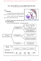
The Cori Cycle
Animals can synthesize glucose 6-phosphate via gluconeogenesis just like all other species. However,
unlike most species, animals can convert glucose 6-phosphate to glucose, which is secreted into the
circulatory system. Mammals, in particular, have a sophisticated cycle of secretion and uptake of
glucose. It's called the Cori cycle after the Nobel Laureates: Carl Ferdinand Cori and Gerty Theresa
Cori.
The glucose 6-phosphate molecules synthesized in the liver can either be converted to glycogen
[Glycogen Synthesis] or converted to glucose and secreted into the blood stream. The glucose molecules
are taken up by muscle cells where they can be stored as glucogen. During strenuous exercise the
glycogen is broken down to glucose 6-phosphate [Glycogen Degradation] and oxdized via the glycolysis
pathway. This pathway yields ATP that is used in muscle contraction.
If oxygen is limiting, the end product of glucose breakdown isn't CO2 but lactate. Lactate is secreted into
the blood stream where it is taken up by the liver and converted to pyruvate by the enzyme lactate
dehydrogenase. Pyruvate is the substrate for gluconeogenesis. The synthesis of glucose in the liver
requires energy in the form of ATP and this energy is supplied by a variety of sources. The breakdown of
fatty acids is the source shown in the figure.
The Cori cycle preserves carbon atoms. The six carbon molecule, glucose, is split into two 3-carbon
molecules (lactate) that are then converted to another 3-carbon molecule (pyruvate). Two pyruvates are
joined to make glucose.
Production of biocellulose (bacterial cellulose)
1. Biocellulose
Cellulose is the main component of plant cell wall. Some bacteria produce cellulose (celled biocellulose
or bacterial cellulose). Plant cellulose and bacterial cellulose have the same chemical structure, but
different physical and chemical properties. Figure 1 shows an electron microscopic image of
biocellulose and plant cellulose. Bacterial cellulose is produced by an acetic acid-producing bacterium,
Acetobacter xylinum. The diameter of biocellulose is about 1/100 of that of plant cellulose and Young's
modulus of biocellulose is almost equivalent to that of aluminum. Therefore, biocellulose is expected to
be a new biodegradable biopolymer.
Fig. 1. Bacterial cellulose and plant cellulose.
2. Production of biocellulose in an airlift reactor
In the mass production of biocellulose, conventionally an agitated reactor is used. In our laboratory, we
applied an airlift reactor to produce bacterial cellulose because this reactor is simple in structure, its
energy requirement is low, its shear stress to cells is small, and the possibility of contamination is low.
Figure 2 shows a 50-liter airlift reactor. In the airlift reactor, the productivity of bacterial cellulose was
equivalent to that in conventional agitated reactors and its energy requirement was one-tenth of that in
agitated reactors. The bacterial cellulose produced in an airlift reactor formed a unique pellet-type
cellulose.
Fig. 2. 50 liter airlift reactor.
3. Analysis of genes for biocellulose synthesis
All genes responsible for biocellulose synthesis have been cloned and their characterization is under
way. Figure 3 shows the predicted steps of bacterial cellulose synthesis when glucose is used as the
carbon source. The analysis of genes will lead to higher productivity of bacterial cellulose and to new
biocellulose with different properties.
Fig. 3. The predicted pathway of cellulose synthesis and secretion when glucose is
taken into Gluconacetobactor xylinum from the outside of the cell.
4. Future aspects
Preservation of forest resources is essential to prevent global warming because the increase in CO 2
concentration can be stopped only by the absorption of CO2 by plants and trees. However, the use of
trees for the production of paper and construction materials has continuously depleated forest resources.
Bacterial cellulose is the only alternative for plant cellulose because bacteria produce bacterial cellulose
in a few days, while trees need more than 30 years to realize full growth. In this respect, bacterial
cellulose is the key material for preventing global warming and preservation of the nature.
Metabolism of Major NonGlucose Sugars
Fructose Metabolism
Diets containing large amounts of sucrose (a disaccharide of glucose and fructose) can utilize
the fructose as a major source of energy. The pathway to utilization of fructose differs in
muscle and liver.
Muscle which contains only hexokinase can phosphorylate fructose to F6P which is a direct
glycolytic intermediate.
In the liver which contains mostly glucokinase, which is specific for glucose as its substrate,
requires the function of additional enzymes to utilize fructose in glycolysis. Hepatic fructose is
phosphorylated on C1 by fructokinase yielding fructose1phosphate (F1P). In liver the form
of aldolase that predominates (aldolase B) can utilize both F1,6BP and F1P as substrates.
Therefore, when presented with F1P the enzyme generates DHAP and glyceraldehyde. The
DHAP is converted, by triose phosphate isomerase, to G3P and enters glycolysis. The
glyceraldehyde can be phosphorylated to G3P by glyceraldehyde kinase or converted to
DHAP through the concerted actions of alcohol dehydrogenase, glycerol kinase and
glycerol phosphate dehydrogenase.
Three inherited abnormalities in fructose metabolism have been identified. Essential
fructosuria is a benign metabolic disorder caused by the lack of fructokinase which is
normally present in the liver, pancreatic islets and kidney cortex. The fructosuria of this disease
depends on the time and amount of fructose and sucrose intake. Since the disorder is
asymptomatic and harmless it may go undiagnosed.
Hereditary fructose intolerance is a potentially lethal disorder resulting from a lack of aldolase
B which is normally present in the liver, small intestine and kidney cortex. The disorder is
characterized by severe hypoglycemia and vomiting following fructose intake. Prolonged intake
of fructose by infants with this defect leads to vomiting, poor feeding, jaundice, hepatomegaly,
hemorrhage and eventually hepatic failure and death. The hypoglycemia that result following
fructose uptake is caused by fructose1phosphate inhibition of glycogenolysis, by interfering
with the phosphorylase reaction, and inhibition of gluconeogenesis at the deficient aldolase
step. Patients remain symptom free on a diet devoid of fructose and sucrose.
Hereditary fructose1,6bisphosphatase deficiency results in severely impaired hepatic
gluconeogenesis and leads to episodes of hypoglycemia, apnea, hyperventillation, ketosis and
lactic acidosis. These symptoms can take on a lethal course in neonates. Later in life episodes
are triggered by fasting and febrile infections.
Clinical Significance of Fructose Metabolism
Galactose Metabolism
Galactose, which is metabolized from the milk sugar, lactose (a disaccharide of glucose and
galactose), enters glycolysis by its conversion to glucose1phosphate (G1P).
This occurs through a series of steps. First the galactose is phosphorylated by galactokinase
to yield galactose1phosphate. Epimerization of galactose1phosphate to G1P requires the
transfer of UDP from uridine diphosphoglucose (UDPglucose) catalyzed by galactose1
phosphate uridyl transferase (official name: UDPglucosehexose1phosphate
uridylyltransferase). This generates UDPgalactose and G1P. The UDPgalactose is
epimerized to UDPglucose by UDPgalactose4 epimerase (see reaction mechanism). The
UDP portion is exchanged for phosphate generating glucose1phosphate which then is
converted to G6P by phosphoglucose mutase.
Galactose on the Web:
Metabolic Pathways of Biochemistry: Galactose Pathway
Clinical Significance of Galactose Metabolism
Three inherited disorders of galactose metabolism have been delineated. Classic galactosemia
is a major symptom of two enzyme defects.One results from loss of the enzyme galactose1
phosphate uridyl transferase.The second form of galactosemia results from a loss of
galactokinase. These two defects are manifest by a failure of neonates to thrive. Vomiting and
diarrhea occur following ingestion of milk, hence individuals are termed lactose intolerant.
Clinical findings of these disorders include impaired liver function (which if left untreated leads
to severe cirrhosis), elevated blood galactose, hypergalactosemia, hyperchloremic metabolic
acidosis, urinary galactitol excretion and hyperaminoaciduria. Unless controlled by exclusion of
galactose from the diet, these galactosemias can go on to produce blindness and fatal liver
damage. Even on a galactoserestricted diet, transferasedeficient individuals exhibit urinary
galacitol excretion and persistently elevated erythrocyte galactose1phosphate levels.
Blindness is due to the conversion of circulating galactose to the sugar alcohol galacitol, by an
NADPHdependent galactose reductase that is present in neural tissue and in the lens of the
eye. At normal circulating levels of galactose this enzyme activity causes no pathological
effects. However, a high concentration of galacitol in the lens causes osmotic swelling, with the
resultant formation of cataracts and other symptoms. The principal treatment of these
disorders is to eliminate lactose from the diet.
The third disorder of galactose metabolism result from a deficiency of UDPgalactose4
epimerase. Two different forms of this deficiency have been found. One is benign affecting
only red and white blood cells. The other affects multiple tissues and manifests symptoms
similar to the transferase deficiency. Treatment involves restriction of dietary galactose.
Mannose Metabolism
The digestion of many polysaccharides and glycoproteins yields mannose which is
phosphorylated by hexokinase to generate mannose6phosphate. Mannose6phosphate is
converted to fructose6phosphate, by the enzyme phosphomannose isomerase, and then
enters the glycolytic pathway or is converted to glucose6phosphate by the gluconeogenic
pathway of hepatocytes.
In eukaryotes,mannose is constituent of N and Olinked glycans as well as GPI anchors.
GDPmannose is the donor form of mannose.
Glycerol Metabolism
The predominant source of glycerol is adipose tissue. This molecule is the backbone for the
triacylglycerols. Following release of the fatty acid portions of triacylglycerols the glycerol
backbone is transported to the liver where it it phosphorylated by glycerol kinase yielding
glycerol3phosphate. Glycerol3phosphate is oxidized to DHAP by glycerol3phosphate
dehydrogenase. DHAP then enters the glycolytic if the liver cell needs energy. However, the
more likely fate of glycerol is to enter the gluconeogenesis pathway in order for the liver to
produce glucose for use by the rest of the body.
Glucuronate Metabolism
Glucuronate is a highly polar molecule which is incorporated into proteoglycans as well as
combining with bilirubin and steroid hormones; it can also be combined with certain drugs to
increase their solubility. Glucuronate is derived from glucose in the uronic acid pathway.
The uronic acid pathway is utilized to synthesize UDPglucuronate, glucuronate and L
ascorbate. The pathway involves the oxidation of glucosae6phosphate to UDPglucuronate.
The oxidation is uncoupled from energy production. UDPglucuronate is used in the synthesis
of glycosaminoglycan and proteoglycans as well as forming complexes with bilirubin, steroids
and certain drugs. The glucuronate complexes form to solubilize compounds for excretion. The
synthesis of ascorbate (vitamin C) does not occur in primates.
The uronic acid pathway is an alternative pathway for the oxidation of glucose that does not
provide a means of producing ATP, but is utilized for the generation of the activated form of
glucuronate, UDPglucuronate. The uronic acid pathway of glucose conversion to glucuronate
begins by conversion of glucose6phosphate is to glucose1phosphate by
phosphoglucomutase, and then activated to UDPglucose by UDPglucose
pyrophosphorylase. UDPglucose is oxidized to UDPglucuronate by the NAD +requiring
enzyme, UDPglucose dehydrogenase. UDPglucuronate then serves as a precursor for the
synthesis of iduronic acid and UDPxylose and is incorporated into proteoglycans and
glycoproteins or forms conjugates with bilirubin, steroids, xenobiotics, drugs and many
compounds containing hydroxyl (OH) groups.
Clinical Significance of Glucuronate
In the adult human, a significant number of erythrocytes die each day. This turnover releases
significant amounts of the ironfree portion of heme, porphyrin, which is subsequently
degraded. The primary sites of porphyrin degradation are found in the reticuloendothelial cells
of the liver, spleen and bone marrow. The breakdown of porphyrin yields bilirubin, a product
that is nonpolar and therefore, insoluble. In the liver, to which is transported in the plasma
bound to albumin, bilirubin is solubilized by conjugation to glucuronate. The soluble conjugated
bilirubin diglucuronide is then secreted into the bile. An inability to conjugate bilirubin, for
instance in hepatic disease or when the level of bilirubin production exceeds the capacity of
the liver, is a contributory cause of jaundice.
The conjugation of glucuronate to certain nonpolar drugs is important for their solubilization in
the liver. Glucuronate conjugated drugs are more easily cleared from the blood by the kidneys
for excretion in the urine. The glucuronatedrug conjugation system can, however, lead to drug
resistance; chronic exposure to certain drugs, such as barbiturates and AZT, leads to an
increase in the synthesis of the UDPglucuronyltransferases in the liver that are involved in
glucuronatedrug conjugation. The increased levels of these hepatic enzymes result in a higher
rate of drug clearance leading to a reduction in the effective dose of glucuronate cleared drugs.
PHOTOSYNTHESIS - - an understandable (not necessarily easy) approach........
Pinus palustris---Pearson Creek
Everything should be made as simple as possible, but not simpler. - Albert Einstein
(it will take some time to understand this; read deliberately and understand what you have read before
going on to the next paragraphs)
Photosynthesis is defined as the formation of carbohydrates in living plants from water and carbon
dioxide (CO2). It is the most important chemical pathway (series of chemical reactions) on our planet.
Almost all of the biomass on Earth was initially created by photosynthesis.
Each year 100 quadrillion (or 10 to the 17th) Kilocalories (K.cal.) of useful energy are produced by
photosynthesis (about 100 times more energy than is consumed by burning of fossil fuels). At least half
of the photosynthesis in the world takes place in oceans, lakes and rivers, brought about by many
different microorganisms that constitute the phytoplankton.
All organisms on Earth can be classified on the basis of two fundamental physiologic requirements:
(A) Energy source:
(1) use sunlight for energy: Phototrophs.
(2)use chemical compounds for energy : Chemotrophs
(B) Carbon source:
(1)source is CO2: Autotrophs.
(2) source is chemical compounds: Heterotrophs
Chemoautotrophs (use chemical compounds for energy and CO2 for carbon)---bacteria (some)
Chemoheterotrophs (use chemical compounds for both energy and carbon)----------animals
Photoaututrophs (use sunlight for energy and CO2 for carbon)-----plants and photosynthetic bacteria
Photoaututrophs utilize sunlight for energy and CO2 for their carbon source by this process of
PHOTOSYNTHESIS whereby sunlight is absorbed by a complex compound known as chlorophyll and
converted to energy which drives a series of chemical reactions that ultimately removes hydrogen from
water or other compounds and then combines the hydrogen with carbon dioxide in a way that produces
sugars.
Photosynthetic organisms can be divided into two classes: those which produce oxygen and those which
do not. Photosynthetic bacteria do not produce oxygen (in fact some of them called anaerobes cannot
tolerate oxygen) and this is considered a more primitive type of photosynthesis (in which the hydrogen
donor is hydrogen sulfide, lactate or other compounds, but not water). Plants and one type of bacteria
(cyanobacteria) do produce oxygen, an evolutionarily more advanced type of photosynthesis (in which
the hydrogen donor is water).
In a broad chemical sense, the opposite of photosynthesis is respiration. Most of life on this planet (all
except in the deep sea vents) depends on the reciprocal photosynthesis-driven production of carbon
containing compounds by a series of reducing (adding electrons) chemical reactions carried out by
plants and then the opposite process of oxidative (removing electrons) chemical reactions by animals
(and plants, which are capable of both photosynthesis and respiration) in which these carbon compounds
are broken down to carbon dioxide and water.
The oxidative chemical reactions of respiration release energy, some of which is heat and some of it is
captured in the form of high energy compunds such as Adenosine triphosphate (ATP) and Nicotinamide
adenide dinucleotide phosphate (NADPH). These compounds have a high energy (unstable) terminal
phosphate bond and that terminal phosphate is easily detached with the transfer of the energy to drive
chemical reactions in the synthesis of other biomolecules. In this case, the ATP loses one phosphate to
become the energy-depleted ADP (Adenosine diphosphate) and the NADPH loses one electron to
become energy-depleted NADP+.
Photosynthesis converts these energy- depleted compounds (ADP and NADP+) back to the high energy
forms (ATP and NADPH) and the energy thus produced in this chemical form is utilized to drive the
chemical reactions necessary for synthesis of sugars and other carbon containing compounds (e.g.,
proteins, fats). The production of high energy ATP and NADPH in plants occurs in what is known as
Light Phase Reactions (Z Scheme) (requires sunlight). The energy releasing reactions which converts
them back to energy-depleted ADP and NADP is known as Dark Phase Reactions (Calvin Cycle) (does
not require light) in which the synthesis of glucose and other carbohydrates occurs.
So we can summarize by saying that the photosynthetic plants trap solar energy to form ATP and
NADPH (Light Phase) and then use these as the energy source to make carbohydrates and other
biomolecules from carbon dioxide and water (Dark Phase), simultaneously releasing oxygen in to the
atmosphere. The chemoheterotrophic animals reverse this process by using the oxygen to degrade the
energy-rich organic products of photosynthesis to CO2 and water in order to generate ATP for their own
synthesis of biomolecules.
Plant photosynthesis, both the Light Phase and Dark phase reactions, takes place in chloroplasts, which
may be regarded as the "power plants" of the green leaf cells. At night, when there is no sunlight energy,
ATP continues to be generated for the plant's needs by respiration, i.e., oxidation of (photosynthetically
produced) carbohydrate in mitochondria (similar to animals).
Chloroplasts have many shapes in different species but are generally fusiform shaped (and much larger
than mitochondria) and have many flattened membrane-surrounded vesicles called thylakoids which are
arranged in stacks called grana. These thylakoid membranes contain all of the photosynthetic pigments
of the chloroplast and all of the enzymes required for Light Phase reactions. The fluid in the stroma
surrounding the thylakoid vesicles contains most of the enzymes for Dark phase reactions.
There are several light-absorbing pigments in the thylakoid membranes. The most important are the
green chlorophylls which are complex protoporphyrin (resembles hemoglobin) molecules which have a
magnesiun ion in the center. There are two types of chlorophyll: chlorophyll a, which is always present
in all green plants, and a second, chlorophyll b which is also present (about half as much as chlorophyll
a) in some plants. The chlorophylls are the major light receptors, absorbing light mostly in the 400 to
500 and 600 to 700 nanometer (nm.)wavelength ranges. The absorption spectra for chlorphylls a and b
are shown below. Other pigmented compounds present in the thylakoid membranes include carotenoids
(are red, yellow or purple), the most important of which is beta-carotene, the precursor of vitamin A in
animals. The carotenoid pigments absorb sunlight at wavelengths other than those absorbed by the
chlorophylls and thus are supplementary light receptors.
The thylakoid membranes of plant chloroplasts have two different sets of light harvesting chlorophyll
and carotenoid molecules combined with a special protein. There are two of these Photochemical
Reaction Centers:
Photosystem I: has a high ratio of chlorophyll a to chlorophyll b.
PhotosystemII: has relatively more chlorophyll b and may also contain a chlorophyll c.
The plants and cyanobacteria (which use water as a hydrogen donor and produce oxygen) have
Photosystems I and II, whereas the less highly evolved other photosynthetic bacteria(which do not use
water as their hydgrogen donor and do not produce oxygen) have only Photosystem I.
How does the absorption of light by the chlorophyll pigments in the thylakoid membrane cause the
conversion of light energy to chemical (ATP & NADHP) energy?
The quick answer is that an electron is stripped from water and transferred to NADP+ to form NADPH
which is an endergonic (requires energy imput) reaction.That energy is supplied by the sunlight
absorbed in the chloroplasts. And in the process, a phosphorus is added to ADP to produce ATP.
When the chlorophyll molecule is excited by light, the energy level of an electron in its structure is
"boosted to a higher energy level and this "excited" chlorophyll (now is called an exciton) moves rapidly
the the reaction center of the Photosystem I where it transfers its extra energy to an electron which is
then expelled from the reaction center and is accepted by the first member of a chain of electron carriers
and ultimately reaches NADP+, reducing it to NADPH. The reaction center has lost an electron and this
"electron hole" is filled by by stripping electrons from water which leaves hydrogen ion (H+) and
molecular oxygen (O2). The pathway of electrons from water to NADP+ has "Z" shape when diagramed
and is refered to as the Z Scheme.
The Z Scheme diagram shows the pathway of an electron from water (lower right) to NADP+ (upper
left). It also shows the energy relationships which are measured as voltage potential shown on the
scaleon the right. To raise the energy of the electrons derived from water (+0.82 volts) to the level
necessary to reduce NADP+ to NADPH (-0.32 volts), each electron must be boosted twice (vertical red
arrows) by light energy absorbed in Photosystems I and II. After each boosting , the energized electrons
flow "downhill" (diagonal black lines) and in the process transfer some of their energy to a series of
reactions which ultimately adds a phosporus to ADP to produce high energy ATP and reduces NADP+ to
NADPH. There is an alternative shunt whereby the electron flow turns back to cytochrome b563 (green
line)and this is called cyclic electron flow and it occurs when there is no need for NADPH, so only ATP
is produced.
How are the electrons lost from Photocenters replaced? The "electron hole" in Photosystem I is filled by
the electron which was expelled by sunlight energy from Photosytem II and travels to Photosystem I via
the chain of electron carriers (the right red vertical and right black diagonal lines). Then the resulting
"electron hole" in Photosystem II is in turn filled by the splitting of water (by an enzyme named water
dehydrogenase) into electrons and H+ ions and molecular oxygen. The electrons go to Photosystem II
"electron holes" and the H+s go into the fluid medium and the oxygen is released into the air.
For each electron flowing from water to NADP+ (a net change in 1.14 volts), two quanta of light are
absorbed, one by each Photosystem. Each molecule of oxygen released involves the flow of four
electrons from two water molecules to two NADP+s and requires four quanta of sunlight absorbed by
each Photosystem to provide the energy to do this. These are the "Light Phase Reactions" of
photosynthesis, which produce two high energy chemical products, namely NADPH and ATP.
Now what are the "Dark Phase Reactions" (aka Calvin Cycle)? This is the cycle that converts CO2 into
glucose. Since it utilizes the chemical energy in the ATP and NADPH, it does not require sunlight (hence
the name). It is a complex cycle of mostly phosphorylation (adding or removing phosphate) and
oxidative (electron removal) chemical reactions whereby 6 molecules of CO2 are converted into one
molecule of glucose. It requires the energy-releasing cleavage of high energy bonds of 18 ATPs and 12
NADPHs . The resulting 18 ADPs and 12 NADP+s are then restored by the Light Phase process to their
high energy forms (ATP and NADPH).
Therefor these two (Light and Dark) phases are interlinked and complimentary. And in the end, the
plants have utilized the energy of sunlight to produce glucose (and ultimately other carbohydrates,
proteins and fats) and oxygen from water and carbon dioxide.
he Molecule of Life
A. Carbohydrates
Carbohydrates are a class of organic macromolecules made up of the so called "sugars and
starches". There are three classes of carbohydrates, based on the number of sugar units:
1) Monosaccharides
2) Polysaccharides
1) Monosaccharides (simple sugars)
These molecules consist of open-chain or ring forms of 3 to 8 carbon atoms. The most
common type of monosaccharide is the simple sugar "glucose".
Glucose is an important energy source in metabolically active cells.
Another important monosaccharides are shown below.
Fructose is a common sugar in fruit), and Galactose is the sugar found in milk.
Sugars with 6 carbons are called "hexoses". Five carbon sugars are "pentoses". Whereas 7
carbon sugars are called "heptoses".
Two very important "pentoses" (5 carbons) are, Ribose found in Ribonucleic Acid, RNA, and
Deoxyribose found in Deoxyribonucleic Acid, DNA.
Disaccharides
When two monosaccharides are joined together they form a "disaccharide".
This linking of two sugars involves the removal of a molecule of H 2O (water) and is therefore
called a "dehydration linkage". The reaction is called "dehydration synthesis".
e.g. Glucose + Glucose = Maltose
This forms a bond between the #1 carbon of one glucose and the #4 carbon of the other,
therefore it is called an 1-4 linkage, (or Glycosidic Linkage).
Other disaccharides are:
Glucose + Fructose = Sucrose
Glucose + Galactose = Lactose
Polysaccharides
These are long chains of monosaccharides linked together by dehydration linkages.
- Xem thêm -